2UZJ | pdb_00002uzj

Crystal structure of the mature streptococcal cysteine protease, mSpeB


Experimental Data Snapshot

  • Method: X-RAY DIFFRACTION
  • Resolution: 1.55 Å
  • R-Value Free: 
    0.227 (Depositor), 0.222 (DCC) 
  • R-Value Work: 
    0.181 (Depositor), 0.176 (DCC) 
  • R-Value Observed: 
    0.183 (Depositor) 

Starting Model: experimental
View more details

wwPDB Validation   3D Report Full Report


Ligand Structure Quality Assessment 


This is version 1.4 of the entry. See complete history


Literature

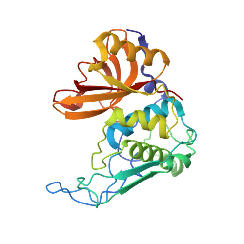
Structure of the Mature Streptococcal Cysteine Protease Exotoxin Mspeb in its Active Dimeric Form.

Olsen, J.G.Dagil, R.Niclasen, L.M.Soerensen, O.E.Kragelund, B.B.

(2009) J Mol Biology 393: 693

  • DOI: https://doi.org/10.1016/j.jmb.2009.08.046
  • Primary Citation of Related Structures:  
    2UZJ

  • PubMed Abstract: 

    Invasive infections of Streptococcus pyogenes are dependent on the cysteine protease streptococcal pyrogenic exotoxin B. Previous structures of the enzyme have not disclosed the proper active-site configuration. Here, the crystal structure of the mature enzyme is presented to 1.55 A, disclosing a homodimer. A serine from one subunit inserts into the active site of the other to donate to the oxyanion hole and coordinates the ligand proximal to the active-site cysteine. Dimerization is unique to the mature form and is clearly a prerequisite for catalysis. The present structure supports a tripartite switch system that is triggered upon dimerization and substrate binding: (1) liberation of the active-site histidine from an inactive configuration, (2) relocation of residues blocking the substrate binding pockets and (3) repositioning of two active-site tryptophans to settle in the active configuration. Based on the present structure, the active site of clan CA cysteine proteases is expanded and a detailed mechanism of the deacylation mechanism is proposed. The results may have applications for the development of protease inhibitors specific to bacterial cysteine proteases.


  • Organizational Affiliation

    Structural Biology and NMR Laboratory, Department of Biology, University of Copenhagen, Ole Maaloes Vej 5, Copenhagen, Denmark.


Macromolecules
Find similar proteins by:  (by identity cutoff)  |  3D Structure
Entity ID: 1
MoleculeChains Sequence LengthOrganismDetailsImage
STREPTOPAIN
A, B
253Streptococcus pyogenesMutation(s): 0 
EC: 3.4.22.10
UniProt
Find proteins for P0C0J0 (Streptococcus pyogenes)
Explore P0C0J0 
Go to UniProtKB:  P0C0J0
Entity Groups  
Sequence Clusters30% Identity50% Identity70% Identity90% Identity95% Identity100% Identity
UniProt GroupP0C0J0
Sequence Annotations
Expand
  • Reference Sequence
Small Molecules
Experimental Data & Validation

Experimental Data

  • Method: X-RAY DIFFRACTION
  • Resolution: 1.55 Å
  • R-Value Free:  0.227 (Depositor), 0.222 (DCC) 
  • R-Value Work:  0.181 (Depositor), 0.176 (DCC) 
  • R-Value Observed: 0.183 (Depositor) 
Space Group: P 1 21 1
Unit Cell:
Length ( Å )Angle ( ˚ )
a = 58.828α = 90
b = 60.888β = 101.14
c = 70.365γ = 90
Software Package:
Software NamePurpose
REFMACrefinement
MOSFLMdata reduction
SCALEPACKdata scaling
CCP4Iphasing

Structure Validation

View Full Validation Report


Ligand Structure Quality Assessment 


Entry History 

Deposition Data

Revision History  (Full details and data files)

  • Version 1.0: 2009-09-08
    Type: Initial release
  • Version 1.1: 2011-05-08
    Changes: Version format compliance
  • Version 1.2: 2011-07-13
    Changes: Version format compliance
  • Version 1.3: 2023-12-13
    Changes: Data collection, Database references, Derived calculations, Other, Refinement description
  • Version 1.4: 2024-10-09
    Changes: Structure summary