3DI3 | pdb_00003di3

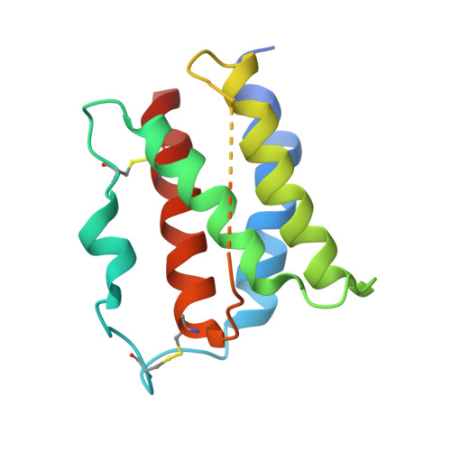
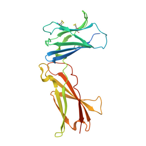
Crystal structure of the complex of human interleukin-7 with glycosylated human interleukin-7 receptor alpha ectodomain


Experimental Data Snapshot

  • Method: X-RAY DIFFRACTION
  • Resolution: 2.90 Å
  • R-Value Free: 
    0.267 (Depositor), 0.260 (DCC) 
  • R-Value Work: 
    0.229 (Depositor), 0.250 (DCC) 
  • R-Value Observed: 
    0.229 (Depositor) 

wwPDB Validation   3D Report Full Report


This is version 2.2 of the entry. See complete history


Literature

Structural and Biophysical Studies of the Human IL-7/IL-7Ralpha Complex.

McElroy, C.A.Dohm, J.A.Walsh, S.T.

(2009) Structure 17: 54-65

  • DOI: https://doi.org/10.1016/j.str.2008.10.019
  • Primary Citation of Related Structures:  
    3DI2, 3DI3

  • PubMed Abstract: 

    IL-7 and IL-7Ralpha bind the gamma(c) receptor, forming a complex crucial to several signaling cascades leading to the development and homeostasis of T and B cells. We report that the IL-7Ralpha ectodomain uses glycosylation to modulate its binding constants to IL-7, unlike the other receptors in the gamma(c) family. IL-7 binds glycosylated IL-7Ralpha 300-fold more tightly than unglycosylated IL-7Ralpha, and the enhanced affinity is attributed primarily to an accelerated on rate. Structural comparison of IL-7 in complex to both forms of IL-7Ralpha reveals that glycosylation does not participate directly in the binding interface. The SCID mutations of IL-7Ralpha locate outside the binding interface with IL-7, suggesting that the expressed mutations cause protein folding defects in IL-7Ralpha. The IL-7/IL-7Ralpha structures provide a window into the molecular recognition events of the IL-7 signaling cascade and provide sites to target for designing new therapeutics to treat IL-7-related diseases.


  • Organizational Affiliation
    • Department of Molecular and Cellular Biochemistry, College of Medicine, Comprehensive Cancer Center, Ohio State University, 467 Hamilton Hall, 1645 Neil Avenue, Columbus, OH 43210, USA.

Macromolecules
Find similar proteins by:  (by identity cutoff)  |  3D Structure
Entity ID: 1
MoleculeChains Sequence LengthOrganismDetailsImage
Interleukin-7154Homo sapiensMutation(s): 1 
Gene Names: IL7
UniProt & NIH Common Fund Data Resources
Find proteins for P13232 (Homo sapiens)
Explore P13232 
Go to UniProtKB:  P13232
PHAROS:  P13232
GTEx:  ENSG00000104432 
Entity Groups  
Sequence Clusters30% Identity50% Identity70% Identity90% Identity95% Identity100% Identity
UniProt GroupP13232
Sequence Annotations
Expand
  • Reference Sequence
Find similar proteins by:  (by identity cutoff)  |  3D Structure
Entity ID: 2
MoleculeChains Sequence LengthOrganismDetailsImage
Interleukin-7 receptor subunit alpha223Homo sapiensMutation(s): 1 
Gene Names: IL7R
UniProt & NIH Common Fund Data Resources
Find proteins for P16871 (Homo sapiens)
Explore P16871 
Go to UniProtKB:  P16871
PHAROS:  P16871
GTEx:  ENSG00000168685 
Entity Groups  
Sequence Clusters30% Identity50% Identity70% Identity90% Identity95% Identity100% Identity
UniProt GroupP16871
Glycosylation
Glycosylation Sites: 3Go to GlyGen: P16871-1
Sequence Annotations
Expand
  • Reference Sequence
Oligosaccharides

Help

Entity ID: 3
MoleculeChains Length2D Diagram Glycosylation3D Interactions
2-acetamido-2-deoxy-beta-D-glucopyranose-(1-4)-2-acetamido-2-deoxy-beta-D-glucopyranose
C, D, E
2N-Glycosylation
Glycosylation Resources
GlyTouCan:  G42666HT
GlyCosmos:  G42666HT
GlyGen:  G42666HT
Experimental Data & Validation

Experimental Data

  • Method: X-RAY DIFFRACTION
  • Resolution: 2.90 Å
  • R-Value Free:  0.267 (Depositor), 0.260 (DCC) 
  • R-Value Work:  0.229 (Depositor), 0.250 (DCC) 
  • R-Value Observed: 0.229 (Depositor) 
Space Group: P 21 21 21
Unit Cell:
Length ( Å )Angle ( ˚ )
a = 55.573α = 90
b = 68.235β = 90
c = 112.273γ = 90
Software Package:
Software NamePurpose
CNSrefinement
PDB_EXTRACTdata extraction
HKL-2000data collection
HKL-2000data reduction
HKL-2000data scaling

Structure Validation

View Full Validation Report


Entry History 

Deposition Data

Revision History  (Full details and data files)

  • Version 1.0: 2009-01-27
    Type: Initial release
  • Version 1.1: 2011-07-13
    Changes: Non-polymer description, Version format compliance
  • Version 2.0: 2020-07-29
    Type: Remediation
    Reason: Carbohydrate remediation
    Changes: Advisory, Atomic model, Data collection, Database references, Derived calculations, Structure summary
  • Version 2.1: 2021-10-20
    Changes: Database references, Structure summary
  • Version 2.2: 2024-10-16
    Changes: Data collection, Structure summary