4ELM | pdb_00004elm

Crystal structure of the mouse CD1d-lysosulfatide-Hy19.3 TCR complex


Experimental Data Snapshot

  • Method: X-RAY DIFFRACTION
  • Resolution: 3.48 Å
  • R-Value Free: 
    0.265 (Depositor), 0.270 (DCC) 
  • R-Value Work: 
    0.209 (Depositor), 0.210 (DCC) 
  • R-Value Observed: 
    0.212 (Depositor) 

wwPDB Validation   3D Report Full Report


Ligand Structure Quality Assessment 

Created with Raphaël 2.3.0Worse 01 BetterLigand structure goodness of fit to experimental dataBest fitted SGFClick on this verticalbar to view detailsBest fitted PLMClick on this verticalbar to view detailsBest fitted NAGClick on this verticalbar to view details

This is version 2.2 of the entry. See complete history


Literature

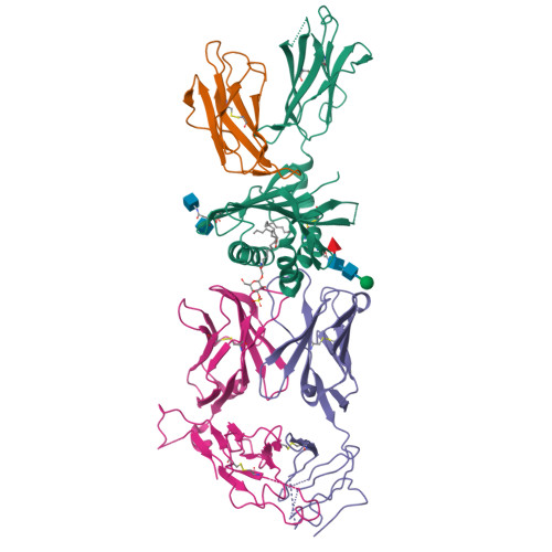
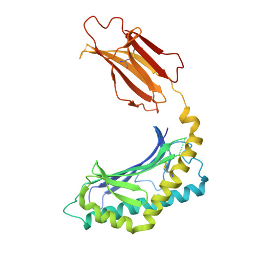
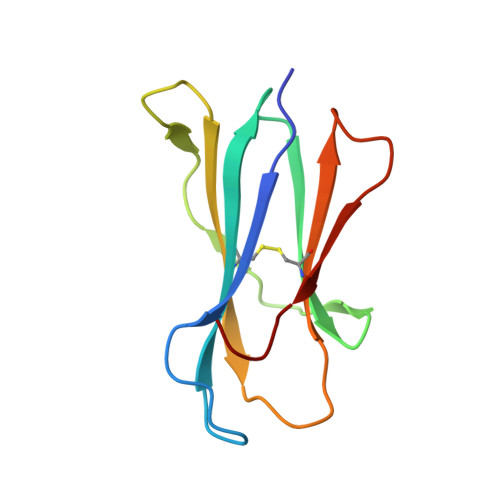
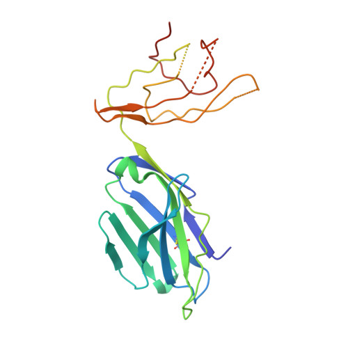
Type II natural killer T cells use features of both innate-like and conventional T cells to recognize sulfatide self antigens.

Girardi, E.Maricic, I.Wang, J.Mac, T.T.Iyer, P.Kumar, V.Zajonc, D.M.

(2012) Nat Immunol 13: 851-856

  • DOI: https://doi.org/10.1038/ni.2371
  • Primary Citation of Related Structures:  
    4ELK, 4ELM

  • PubMed Abstract: 

    Glycolipids presented by the major histocompatibility complex (MHC) class I homolog CD1d are recognized by natural killer T cells (NKT cells) characterized by either a semi-invariant T cell antigen receptor (TCR) repertoire (type I NKT cells or iNKT cells) or a relatively variable TCR repertoire (type II NKT cells). Here we describe the structure of a type II NKT cell TCR in complex with CD1d-lysosulfatide. Both TCR α-chains and TCR β-chains made contact with the CD1d molecule with a diagonal footprint, typical of MHC-TCR interactions, whereas the antigen was recognized exclusively with a single TCR chain, similar to the iNKT cell TCR. Type II NKT cell TCRs, therefore, recognize CD1d-sulfatide complexes by a distinct recognition mechanism characterized by the TCR-binding features of both iNKT cells and conventional peptide-reactive T cells.


  • Organizational Affiliation
    • Division of Cell Biology, La Jolla Institute for Allergy & Immunology, La Jolla, California, USA.

Macromolecules
Find similar proteins by:  (by identity cutoff)  |  3D Structure
Entity ID: 1
MoleculeChains Sequence LengthOrganismDetailsImage
Antigen-presenting glycoprotein CD1d1A,
B [auth C]
285Mus musculusMutation(s): 0 
Gene Names: Cd1.1CD1dCd1d1
UniProt & NIH Common Fund Data Resources
Find proteins for P11609 (Mus musculus)
Explore P11609 
Go to UniProtKB:  P11609
IMPC:  MGI:107674
Entity Groups  
Sequence Clusters30% Identity50% Identity70% Identity90% Identity95% Identity100% Identity
UniProt GroupP11609
Glycosylation
Glycosylation Sites: 3Go to GlyGen: P11609-1
Sequence Annotations
Expand
  • Reference Sequence
Find similar proteins by:  (by identity cutoff)  |  3D Structure
Entity ID: 2
MoleculeChains Sequence LengthOrganismDetailsImage
Beta-2 microglobulinC [auth B],
D
99Mus musculusMutation(s): 0 
Gene Names: B2mbeta-2-microglobulinmCG_11606RP23-34E24.5-001
UniProt & NIH Common Fund Data Resources
Find proteins for P01887 (Mus musculus)
Explore P01887 
Go to UniProtKB:  P01887
IMPC:  MGI:88127
Entity Groups  
Sequence Clusters30% Identity50% Identity70% Identity90% Identity95% Identity100% Identity
UniProt GroupP01887
Sequence Annotations
Expand
  • Reference Sequence
Find similar proteins by:  (by identity cutoff)  |  3D Structure
Entity ID: 3
MoleculeChains Sequence LengthOrganismDetailsImage
Hy19.3 TCR alpha chain (mouse variable domain, human constant domain)
E, G
208Mus musculusMutation(s): 0 
Gene Names: Valpha1 (mouse variable domainhuman constant domain
Entity Groups  
Sequence Clusters30% Identity50% Identity70% Identity90% Identity95% Identity100% Identity
Sequence Annotations
Expand
  • Reference Sequence
Find similar proteins by:  (by identity cutoff)  |  3D Structure
Entity ID: 4
MoleculeChains Sequence LengthOrganismDetailsImage
Hy19.3 TCR beta chain (mouse variable domain, human constant domain)
F, H
244Mus musculusMutation(s): 0 
Gene Names: Vbeta16 (mouse variable domainhuman constant domain)
Entity Groups  
Sequence Clusters30% Identity50% Identity70% Identity90% Identity95% Identity100% Identity
Sequence Annotations
Expand
  • Reference Sequence
Oligosaccharides

Help

Entity ID: 5
MoleculeChains Length2D Diagram Glycosylation3D Interactions
2-acetamido-2-deoxy-beta-D-glucopyranose-(1-4)-2-acetamido-2-deoxy-beta-D-glucopyranose
I
2N-Glycosylation
Glycosylation Resources
GlyTouCan:  G42666HT
GlyCosmos:  G42666HT
GlyGen:  G42666HT
Entity ID: 6
MoleculeChains Length2D Diagram Glycosylation3D Interactions
beta-D-mannopyranose-(1-4)-2-acetamido-2-deoxy-beta-D-glucopyranose-(1-4)-[alpha-L-fucopyranose-(1-6)]2-acetamido-2-deoxy-beta-D-glucopyranose
J
4N-Glycosylation
Glycosylation Resources
GlyTouCan:  G32152BH
GlyCosmos:  G32152BH
GlyGen:  G32152BH
Small Molecules
Ligands 3 Unique
IDChains Name / Formula / InChI Key2D Diagram3D Interactions
SGF
Query on SGF

Download Ideal Coordinates CCD File 
M [auth A],
Q [auth C]
(2S,3R,4E)-2-amino-3-hydroxyoctadec-4-en-1-yl 3-O-sulfo-beta-D-galactopyranoside
C24 H47 N O10 S
BXSULSOCJNTUJS-YTBMLWRQSA-N
PLM
Query on PLM

Download Ideal Coordinates CCD File 
N [auth A],
R [auth C]
PALMITIC ACID
C16 H32 O2
IPCSVZSSVZVIGE-UHFFFAOYSA-N
NAG
Query on NAG

Download Ideal Coordinates CCD File 
K [auth A],
L [auth A],
O [auth C],
P [auth C]
2-acetamido-2-deoxy-beta-D-glucopyranose
C8 H15 N O6
OVRNDRQMDRJTHS-FMDGEEDCSA-N
Experimental Data & Validation

Experimental Data

  • Method: X-RAY DIFFRACTION
  • Resolution: 3.48 Å
  • R-Value Free:  0.265 (Depositor), 0.270 (DCC) 
  • R-Value Work:  0.209 (Depositor), 0.210 (DCC) 
  • R-Value Observed: 0.212 (Depositor) 
Space Group: P 1 21 1
Unit Cell:
Length ( Å )Angle ( ˚ )
a = 98.514α = 90
b = 126.996β = 110.53
c = 104.349γ = 90
Software Package:
Software NamePurpose
PHASERphasing
REFMACrefinement
MOSFLMdata reduction
SCALAdata scaling

Structure Validation

View Full Validation Report


Ligand Structure Quality Assessment 

Created with Raphaël 2.3.0Worse 01 BetterLigand structure goodness of fit to experimental dataBest fitted SGFClick on this verticalbar to view detailsBest fitted PLMClick on this verticalbar to view detailsBest fitted NAGClick on this verticalbar to view details

Entry History 

Deposition Data

Revision History  (Full details and data files)

  • Version 1.0: 2012-07-25
    Type: Initial release
  • Version 1.1: 2013-01-09
    Changes: Database references
  • Version 1.2: 2013-02-06
    Changes: Non-polymer description
  • Version 1.3: 2015-08-12
    Changes: Refinement description
  • Version 2.0: 2020-07-29
    Type: Remediation
    Reason: Carbohydrate remediation
    Changes: Advisory, Atomic model, Data collection, Database references, Derived calculations, Structure summary
  • Version 2.1: 2023-12-27
    Changes: Data collection, Database references, Derived calculations, Structure summary
  • Version 2.2: 2024-11-06
    Changes: Structure summary