7LHK | pdb_00007lhk

High-Resolution Crystal Structure of a Lipin/Pah Phosphatidic Acid Phosphatase


Experimental Data Snapshot

  • Method: X-RAY DIFFRACTION
  • Resolution: 1.95 Å
  • R-Value Free: 
    0.211 (Depositor), 0.210 (DCC) 
  • R-Value Work: 
    0.165 (Depositor), 0.170 (DCC) 
  • R-Value Observed: 
    0.167 (Depositor) 

Starting Model: experimental
View more details

wwPDB Validation   3D Report Full Report


This is version 1.2 of the entry. See complete history


Literature

Structures of a lipin/Pah phosphatidic acid phosphatase in distinct catalytic states reveal a signature motif for substrate recognition.

Vitkovska, T.Welcome, F.S.Khayyo, V.I.Gao, S.Wymore, T.Airola, M.V.

(2025) J Biological Chem : 110830-110830

  • DOI: https://doi.org/10.1016/j.jbc.2025.110830
  • Primary Citation of Related Structures:  
    7LHK, 9D13, 9D14, 9D15, 9D16

  • PubMed Abstract: 

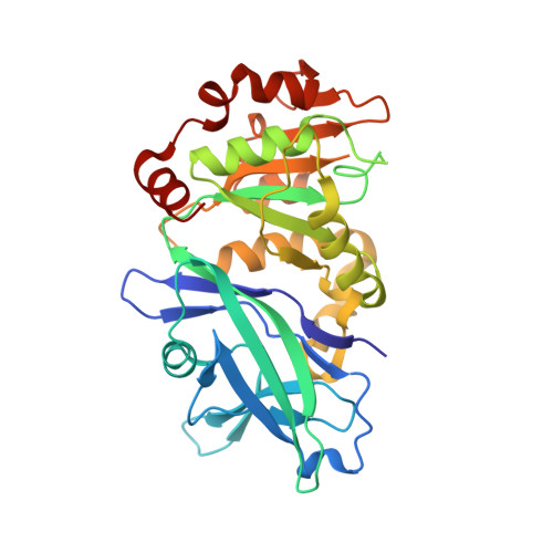
    Lipin/Pah phosphatidic acid phosphatases (PAPs) are Mg 2+ -dependent enzymes that catalyze the dephosphorylation of phosphatidic acid (PA) to produce diacylglycerol. Deficiency of lipin PAP activity in humans results in inflammatory disorders such as rhabdomyolysis and Majeed syndrome. Previously, we reported the first PAP enzyme structures of Tetrahymena thermophila (Tt) Pah2 at 3.0Å resolution. Here, we present five new higher resolution (1.95-2.40Å) structures of Tt Pah2 that represent active states of catalysis, including the product analog tungstate bound to the active site, and an inactive state with a distorted active site. The structures, in conjunction with flexible docking simulations and biochemical analysis, connect two highly conserved aspartate and arginine residues in magnesium coordination and recognition of the substrate PA. Overall, this provides a structural basis for catalysis and defines a signature Asp-Arg motif in lipin/Pah PAPs that enables recognition of their lipid substrate PA, providing insight into how the HAD domain of lipin/Pah PAPs evolved to act on a membrane embedded substrate.


  • Organizational Affiliation
    • Department of Biochemistry and Cell Biology, Stony Brook University, Stony Brook NY 11794, USA.

Macromolecules
Find similar proteins by:  (by identity cutoff)  |  3D Structure
Entity ID: 1
MoleculeChains Sequence LengthOrganismDetailsImage
Nuclear elongation and deformation protein
A, B
313Tetrahymena thermophila SB210Mutation(s): 0 
Gene Names: TTHERM_00215970
UniProt
Find proteins for I7MFJ3 (Tetrahymena thermophila (strain SB210))
Explore I7MFJ3 
Go to UniProtKB:  I7MFJ3
Entity Groups  
Sequence Clusters30% Identity50% Identity70% Identity90% Identity95% Identity100% Identity
UniProt GroupI7MFJ3
Sequence Annotations
Expand
  • Reference Sequence
Experimental Data & Validation

Experimental Data

  • Method: X-RAY DIFFRACTION
  • Resolution: 1.95 Å
  • R-Value Free:  0.211 (Depositor), 0.210 (DCC) 
  • R-Value Work:  0.165 (Depositor), 0.170 (DCC) 
  • R-Value Observed: 0.167 (Depositor) 
Space Group: C 2 2 21
Unit Cell:
Length ( Å )Angle ( ˚ )
a = 144.41α = 90
b = 171.626β = 90
c = 62.845γ = 90
Software Package:
Software NamePurpose
PHENIXrefinement
Aimlessdata scaling
PDB_EXTRACTdata extraction
DIALSdata reduction

Structure Validation

View Full Validation Report


Entry History & Funding Information

Deposition Data


Funding OrganizationLocationGrant Number
American Heart AssociationUnited States19PRE34450192
National Institutes of Health/National Institute of General Medical Sciences (NIH/NIGMS)United StatesR35GM128666
American Heart AssociationUnited States17SDG33410860

Revision History  (Full details and data files)

  • Version 1.0: 2022-02-23
    Type: Initial release
  • Version 1.1: 2023-10-18
    Changes: Data collection, Refinement description
  • Version 1.2: 2025-11-12
    Changes: Database references, Structure summary