7C5F | pdb_00007c5f

Crystal Structure of Glyceraldehyde-3-phosphate dehydrogenase1 from Escherichia coli at 1.88 Angstrom resolution


Experimental Data Snapshot

  • Method: X-RAY DIFFRACTION
  • Resolution: 1.88 Å
  • R-Value Free: 
    0.246 (Depositor), 0.250 (DCC) 
  • R-Value Work: 
    0.180 (Depositor), 0.200 (DCC) 

Starting Model: experimental
View more details

wwPDB Validation   3D Report Full Report


Ligand Structure Quality Assessment 


This is version 1.1 of the entry. See complete history


Literature

Characterization and structure of glyceraldehyde-3-phosphate dehydrogenase type 1 from Escherichia coli.

Zhang, L.Liu, M.R.Yao, Y.C.Bostrom, I.K.Wang, Y.D.Chen, A.Q.Li, J.X.Gu, S.H.Ji, C.N.

(2020) Acta Crystallogr F Struct Biol Commun 76: 406-413

  • DOI: https://doi.org/10.1107/S2053230X20010067
  • Primary Citation of Related Structures:  
    7C5F

  • PubMed Abstract: 

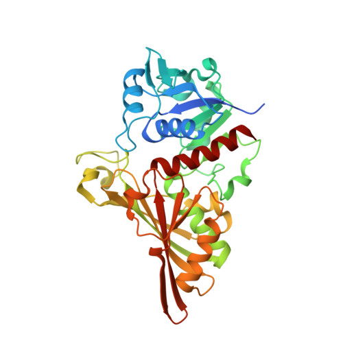
    Glyceraldehyde-3-phosphate dehydrogenase (GAPDH) is a key enzyme in the glycolytic pathway that catalyzes the conversion of D-glyceraldehyde 3-phosphate to 1,3-diphosphoglycerate. Here, the full-length GAPDH type 1 from Escherichia coli (EcGAPDH1) was cloned and overexpressed, and the protein was purified. Biochemical analyses found that the optimum reaction temperature and pH of EcGAPDH1 were 55°C and 10.0, respectively. The protein has a certain amount of thermostability. Crystals of EcGAPDH1 were obtained using the sitting-drop vapor-diffusion technique and X-ray diffraction data were collected to 1.88 Å resolution. Characterization of the crystals showed that they belonged to space group P4 1 2 1 2, with unit-cell parameters a = b = 89.651, c = 341.007 Å, α = β = γ = 90°. The structure of EcGAPDH1 contains four subunits, each of which includes an N-terminal NAD + -binding domain and a C-terminal catalytic domain. Analysis of the NAD + -bound form showed some differences between the structures of EcGAPDH1 and human GAPDH. As EcGAPDH1 shares 100% identity with GAPDH from Shigella sonnei, its structure may help in finding a drug for the treatment of shigellosis.


  • Organizational Affiliation
    • State Key Laboratory of Genetic Engineering, Institute of Genetics, School of Life Sciences, Fudan University, 2005 Songhu Road, Shanghai 200438, People's Republic of China.

Macromolecules
Find similar proteins by:  (by identity cutoff)  |  3D Structure
Entity ID: 1
MoleculeChains Sequence LengthOrganismDetailsImage
Glyceraldehyde-3-phosphate dehydrogenaseA [auth O],
B [auth P],
C [auth Q],
D [auth R]
352Escherichia coli BL21(DE3)Mutation(s): 0 
Gene Names: ECBD_2224
EC: 1.2.1
Entity Groups  
Sequence Clusters30% Identity50% Identity70% Identity90% Identity95% Identity100% Identity
Sequence Annotations
Expand
  • Reference Sequence
Experimental Data & Validation

Experimental Data

  • Method: X-RAY DIFFRACTION
  • Resolution: 1.88 Å
  • R-Value Free:  0.246 (Depositor), 0.250 (DCC) 
  • R-Value Work:  0.180 (Depositor), 0.200 (DCC) 
Space Group: P 41 21 2
Unit Cell:
Length ( Å )Angle ( ˚ )
a = 89.651α = 90
b = 89.651β = 90
c = 341.007γ = 90
Software Package:
Software NamePurpose
REFMACrefinement
HKL-2000data reduction
HKL-2000data scaling
PHASERphasing

Structure Validation

View Full Validation Report


Ligand Structure Quality Assessment 


Entry History & Funding Information

Deposition Data


Funding OrganizationLocationGrant Number
National Key Research and Development Program of ChinaChina2016YFA0500600
Science and Technology Research Program of ShanghaiChina19DZ2282100

Revision History  (Full details and data files)

  • Version 1.0: 2020-09-23
    Type: Initial release
  • Version 1.1: 2023-11-29
    Changes: Data collection, Database references, Refinement description