7LXI | pdb_00007lxi

Crystal structure of S-adenosylmethionine-dependent methyltransferase UmaA from Mycobacterium tuberculosis in complex with SAH


Experimental Data Snapshot

  • Method: X-RAY DIFFRACTION
  • Resolution: 1.95 Å
  • R-Value Free: 
    0.196 (Depositor), 0.197 (DCC) 
  • R-Value Work: 
    0.165 (Depositor), 0.166 (DCC) 
  • R-Value Observed: 
    0.168 (Depositor) 

Starting Model: experimental
View more details

wwPDB Validation   3D Report Full Report


Ligand Structure Quality Assessment 


This is version 1.2 of the entry. See complete history


Literature

Crystal structure of the S-adenosylmethionine-dependent mycolic acid synthase UmaA from Mycobacterium tuberculosis.

Teng, S.Wang, J.Sroge, C.D.Abendroth, J.Lorimer, D.D.Horanyi, P.S.Edwards, T.E.Tillery, L.Craig, J.K.Van Voorhis, W.C.Myler, P.J.Smith, C.L.

(2025) Acta Crystallogr F Struct Biol Commun 81: 146-154

  • DOI: https://doi.org/10.1107/S2053230X25001530
  • Primary Citation of Related Structures:  
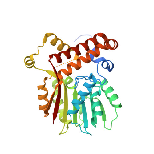
    7LXI

  • PubMed Abstract: 

    Mycobacterium tuberculosis is a Gram-positive bacillus that causes tuberculosis and is a leading cause of mortality worldwide. This disease is a growing health threat due to the occurrence of multidrug resistance. Mycolic acids are essential for generating cell walls and their modification is important to the virulence and persistence of M. tuberculosis. A family of S-adenosylmethionine-dependent mycolic acid synthases modify mycolic acids and represent promising drug targets. UmaA is currently the least-understood member of this family. This paper describes the crystal structure of UmaA. UmaA is a monomer composed of two domains: a structurally conserved SAM-binding domain and a variable substrate-binding auxiliary domain. Fortuitously, our structure contains a nitrate in the active site, a structural mimic of carbonate, which is a known general base in cyclopropane-adding synthases. Further investigation indicated that the structure of the N-terminus is highly flexible. Finally, we have identified S-adenosyl-N-decyl-aminoethyl as a promising potential inhibitor.


  • Organizational Affiliation

    Department of Biology, Washington University in St Louis, St Louis, MO 63134, USA.


Macromolecules
Find similar proteins by:  (by identity cutoff)  |  3D Structure
Entity ID: 1
MoleculeChains Sequence LengthOrganismDetailsImage
S-adenosylmethionine-dependent methyltransferase UmaA294Mycobacterium tuberculosis H37RvMutation(s): 0 
Gene Names: umaARv0469LH57_02505
EC: 2.1.1
UniProt
Find proteins for Q6MX39 (Mycobacterium tuberculosis (strain ATCC 25618 / H37Rv))
Explore Q6MX39 
Go to UniProtKB:  Q6MX39
Entity Groups  
Sequence Clusters30% Identity50% Identity70% Identity90% Identity95% Identity100% Identity
UniProt GroupQ6MX39
Sequence Annotations
Expand
  • Reference Sequence
Small Molecules
Ligands 6 Unique
IDChains Name / Formula / InChI Key2D Diagram3D Interactions
SAH (Subject of Investigation/LOI)
Query on SAH

Download Ideal Coordinates CCD File 
G [auth A]S-ADENOSYL-L-HOMOCYSTEINE
C14 H20 N6 O5 S
ZJUKTBDSGOFHSH-WFMPWKQPSA-N
P6G
Query on P6G

Download Ideal Coordinates CCD File 
B [auth A]HEXAETHYLENE GLYCOL
C12 H26 O7
IIRDTKBZINWQAW-UHFFFAOYSA-N
PO4
Query on PO4

Download Ideal Coordinates CCD File 
E [auth A]PHOSPHATE ION
O4 P
NBIIXXVUZAFLBC-UHFFFAOYSA-K
EDO
Query on EDO

Download Ideal Coordinates CCD File 
D [auth A]1,2-ETHANEDIOL
C2 H6 O2
LYCAIKOWRPUZTN-UHFFFAOYSA-N
NO3
Query on NO3

Download Ideal Coordinates CCD File 
C [auth A],
F [auth A]
NITRATE ION
N O3
NHNBFGGVMKEFGY-UHFFFAOYSA-N
CL
Query on CL

Download Ideal Coordinates CCD File 
H [auth A]CHLORIDE ION
Cl
VEXZGXHMUGYJMC-UHFFFAOYSA-M
Experimental Data & Validation

Experimental Data

  • Method: X-RAY DIFFRACTION
  • Resolution: 1.95 Å
  • R-Value Free:  0.196 (Depositor), 0.197 (DCC) 
  • R-Value Work:  0.165 (Depositor), 0.166 (DCC) 
  • R-Value Observed: 0.168 (Depositor) 
Space Group: P 32 2 1
Unit Cell:
Length ( Å )Angle ( ˚ )
a = 103.22α = 90
b = 103.22β = 90
c = 49.15γ = 120
Software Package:
Software NamePurpose
XDSdata reduction
XSCALEdata scaling
PHENIXrefinement
PDB_EXTRACTdata extraction
PHASERphasing

Structure Validation

View Full Validation Report


Ligand Structure Quality Assessment 


Entry History 

Deposition Data

Revision History  (Full details and data files)

  • Version 1.0: 2021-03-17
    Type: Initial release
  • Version 1.1: 2023-10-18
    Changes: Data collection, Database references, Refinement description
  • Version 1.2: 2025-04-09
    Changes: Data collection, Database references, Structure summary