8CKH | pdb_00008ckh

Crystal structure of IspE from Klebsiella pneumoniae


Experimental Data Snapshot

  • Method: X-RAY DIFFRACTION
  • Resolution: 1.80 Å
  • R-Value Free: 
    0.227 (Depositor), 0.230 (DCC) 
  • R-Value Work: 
    0.188 (Depositor), 0.190 (DCC) 
  • R-Value Observed: 
    0.190 (Depositor) 

Starting Model: experimental
View more details

wwPDB Validation   3D Report Full Report


Ligand Structure Quality Assessment 


This is version 1.1 of the entry. See complete history


Literature

IspE kinase as an anti-infective target: Role of a hydrophobic pocket in inhibitor binding.

Hamid, R.Walsh, D.J.Diamanti, E.Aguilar, D.Lacour, A.Hamed, M.M.Hirsch, A.K.H.

(2024) Structure 32: 2390-2398.e2

  • DOI: https://doi.org/10.1016/j.str.2024.10.009
  • Primary Citation of Related Structures:  
    8CKH, 8QC7, 8QCC, 8QCN, 8QCO

  • PubMed Abstract: 

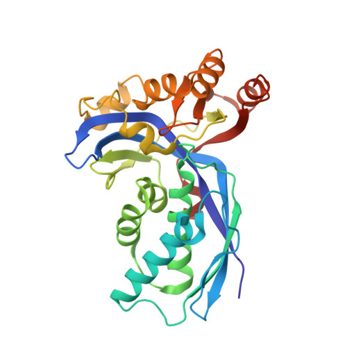
    Enzymes of the methylerythritol phosphate (MEP) pathway are potential targets for antimicrobial drug discovery. Here, we focus on 4-diphosphocytidyl-2-C-methyl-D-erythritol (IspE) kinase from the MEP pathway. We use biochemical and structural biology methods to investigate homologs from pathogenic microorganisms; Escherichia coli, Klebsiella pneumoniae, and Acinetobacter baumannii. We determined the X-ray crystal structures of IspE-inhibitor complexes and studied inhibitors' binding modes targeting the substrate pocket. The experimental results indicate the need for distinct inhibitor strategies due to structural differences among IspE homologs, particularly for A. baumannii IspE, which displays a unique inhibitory profile due to a tighter hydrophobic subpocket in the substrate binding site. This study enhances our understanding of the MEP enzymes and sets the stage for structure-based drug design of selective inhibitors to combat pathogenic microorganisms.


  • Organizational Affiliation

    Helmholtz Institute for Pharmaceutical Research Saarland (HIPS) - Helmholtz Centre for Infection Research (HZI), Campus Building E8.1, 66123 Saarbrücken, Germany; Department of Pharmacy, Saarland University, 66123 Saarbrücken, Germany.


Macromolecules
Find similar proteins by:  (by identity cutoff)  |  3D Structure
Entity ID: 1
MoleculeChains Sequence LengthOrganismDetailsImage
4-diphosphocytidyl-2-C-methyl-D-erythritol kinase
A, B
284Klebsiella pneumoniaeMutation(s): 0 
Gene Names: 
EC: 2.7.1.148
UniProt
Find proteins for A6TAP2 (Klebsiella pneumoniae subsp. pneumoniae (strain ATCC 700721 / MGH 78578))
Explore A6TAP2 
Go to UniProtKB:  A6TAP2
Entity Groups  
Sequence Clusters30% Identity50% Identity70% Identity90% Identity95% Identity100% Identity
UniProt GroupA6TAP2
Sequence Annotations
Expand
  • Reference Sequence
Experimental Data & Validation

Experimental Data

  • Method: X-RAY DIFFRACTION
  • Resolution: 1.80 Å
  • R-Value Free:  0.227 (Depositor), 0.230 (DCC) 
  • R-Value Work:  0.188 (Depositor), 0.190 (DCC) 
  • R-Value Observed: 0.190 (Depositor) 
Space Group: P 21 21 21
Unit Cell:
Length ( Å )Angle ( ˚ )
a = 65.87α = 90
b = 76.781β = 90
c = 98.517γ = 90
Software Package:
Software NamePurpose
PHENIXphasing
PHENIXrefinement
Aimlessdata scaling

Structure Validation

View Full Validation Report


Ligand Structure Quality Assessment 


Entry History & Funding Information

Deposition Data

  • Released Date: 2024-02-28 
  • Deposition Author(s): Hamid, R.

Funding OrganizationLocationGrant Number
Helmholtz AssociationGermany--

Revision History  (Full details and data files)

  • Version 1.0: 2024-02-28
    Type: Initial release
  • Version 1.1: 2025-03-26
    Changes: Database references, Structure summary