8JCR | pdb_00008jcr

Crystal structure of Calotropain FI from Calotropis gigantea (pH 6.0)


Experimental Data Snapshot

  • Method: X-RAY DIFFRACTION
  • Resolution: 1.40 Å
  • R-Value Free: 
    0.175 (Depositor), 0.172 (DCC) 
  • R-Value Work: 
    0.158 (Depositor), 0.155 (DCC) 
  • R-Value Observed: 
    0.158 (Depositor) 

Starting Model: experimental
View more details

wwPDB Validation   3D Report Full Report


Ligand Structure Quality Assessment 


This is version 2.2 of the entry. See complete history


Literature

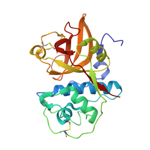
Crystal Structure of Papain-Like Cysteine Protease, Calotropain FI, Purified from the Latex of Calotropis gigantea.

Jamdar, S.N.Kumar, A.Srivastava, G.Makde, R.D.

(2025) J Agric Food Chem 73: 9398-9407

  • DOI: https://doi.org/10.1021/acs.jafc.4c10898
  • Primary Citation of Related Structures:  
    8JCQ, 8JCR

  • PubMed Abstract: 

    The latex of Calotropis gigantea and Calotropis procera elute as two distinct peaks on cation exchange chromatography. One of the peaks is reported to possess multiple papain-like cysteine proteases with different biochemical properties that have been identified with different names. This is mainly due to the absence of a primary sequence for the proteases. Here, we report the crystal structures of calotropain FI from C. gigantea bound to the inhibitor E64 at pH 6 and 9 at 1.25 and 1.4 Å, respectively. Both structures are identical and are very similar to ervatamin B and papain structures. The high quality of electron density maps revealed the primary sequence of calotropain FI. The sequence comparison shows that calotropain FI from C. gigantea is orthologous to procerain, CpCp1-3, and SnuCalCp03 from C. procera and rather distinct from procerain B.


  • Organizational Affiliation

    Food Technology Division, Bhabha Atomic Research Centre, Mumbai 400085, India.


Macromolecules
Find similar proteins by:  (by identity cutoff)  |  3D Structure
Entity ID: 1
MoleculeChains Sequence LengthOrganismDetailsImage
Procerain, Calotropain FI214Calotropis giganteaMutation(s): 0 
Entity Groups  
Sequence Clusters30% Identity50% Identity70% Identity90% Identity95% Identity100% Identity
Sequence Annotations
Expand
  • Reference Sequence
Small Molecules
Modified Residues  1 Unique
IDChains TypeFormula2D DiagramParent
CME
Query on CME
A
L-PEPTIDE LINKINGC5 H11 N O3 S2CYS
Experimental Data & Validation

Experimental Data

  • Method: X-RAY DIFFRACTION
  • Resolution: 1.40 Å
  • R-Value Free:  0.175 (Depositor), 0.172 (DCC) 
  • R-Value Work:  0.158 (Depositor), 0.155 (DCC) 
  • R-Value Observed: 0.158 (Depositor) 
Space Group: C 2 2 21
Unit Cell:
Length ( Å )Angle ( ˚ )
a = 68.675α = 90
b = 120.98β = 90
c = 61.024γ = 90
Software Package:
Software NamePurpose
PHENIXrefinement
XDSdata reduction
Aimlessdata scaling
PHENIXphasing
PHENIXmodel building
Cootmodel building

Structure Validation

View Full Validation Report


Ligand Structure Quality Assessment 


Entry History & Funding Information

Deposition Data


Funding OrganizationLocationGrant Number
Other governmentIndia--

Revision History  (Full details and data files)

  • Version 1.0: 2024-05-22
    Type: Initial release
  • Version 2.0: 2024-11-20
    Type: Coordinate replacement
    Reason: Sequence discrepancy
    Changes: Advisory, Atomic model, Data collection, Database references, Derived calculations, Non-polymer description, Polymer sequence, Refinement description, Structure summary
  • Version 2.1: 2025-03-19
    Changes: Database references
  • Version 2.2: 2025-04-23
    Changes: Database references