8K0W | pdb_00008k0w

Crystal structure of theophylline DNA aptamer bound to theophylline, soaked in Selenourea

  • Classification: DNA
  • Organism(s): synthetic construct
  • Mutation(s): No 

  • Deposited: 2023-07-10 Released: 2025-04-16 
  • Deposition Author(s): Huang, Y., Lin, X., Huang, L.
  • Funding Organization(s): National Natural Science Foundation of China (NSFC)

Experimental Data Snapshot

  • Method: X-RAY DIFFRACTION
  • Resolution: 1.42 Å
  • R-Value Free: 
    0.222 (Depositor), 0.214 (DCC) 
  • R-Value Work: 
    0.208 (Depositor), 0.208 (DCC) 
  • R-Value Observed: 
    0.209 (Depositor) 

Starting Model: experimental
View more details

wwPDB Validation   3D Report Full Report


Ligand Structure Quality Assessment 


This is version 1.0 of the entry. See complete history


Literature

From Theophylline to Adenine or preQ 1 : Repurposing a DNA Aptamer Revealed by Crystal Structure Analysis.

Lin, X.Huang, Y.Huang, J.Yuan, H.Luo, Y.Lu, Z.Ao, Y.Huang, J.Chen, S.B.Miao, Z.Huang, L.

(2025) Angew Chem Int Ed Engl : e202504107-e202504107

  • DOI: https://doi.org/10.1002/anie.202504107
  • Primary Citation of Related Structures:  
    8K0T, 8K0U, 8K0V, 8K0W, 8ZAS

  • PubMed Abstract: 

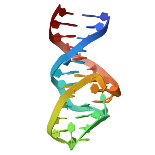
    Aptamers, which are short, single-stranded DNA or RNA, are capable of binding to a wide array of targets with exceptional selectivity. Those with high affinity for theophylline have the potential to serve as biosensors, crucial for tracking theophylline levels in the treatment of respiratory conditions. Despite the extensive structural characterization of the RNA theophylline aptamer, the DNA counterpart's ligand-recognition mechanism has remained unclear. Here, we elucidate the DNA theophylline aptamer's ligand-binding mechanism through high-resolution crystal structures of its complexes with theophylline, 3-methylxanthine, and hypoxanthine. Guided by these structural insights, we computationally redesigned the theophylline-binding pocket via rational mutagenesis of key nucleotides, generating novel aptamers selective for adenine and prequeuosine (preQ 1 ) ligands. These engineered aptamers were validated through biochemical and crystallographic analyses. By integrating structural biology with computational design, our work provides a relatively simple and effective method for developing new aptamers. While this strategy does not supplant SELEX, it serves as a beneficial complement to it.


  • Organizational Affiliation

    Guangdong Provincial Key Laboratory of Malignant Tumor Epigenetics and Gene Regulation, Guangdong-Hong Kong Joint Laboratory for RNA Medicine, Sun Yat-Sen Memorial Hospital, Sun Yat-Sen University, Guangzhou, 510120, China.


Macromolecules
Find similar nucleic acids by:  (by identity cutoff)  |  3D Structure
Entity ID: 1
MoleculeChains LengthOrganismImage
DNA (29-MER)29synthetic construct
Sequence Annotations
Expand
  • Reference Sequence
Small Molecules
Ligands 3 Unique
IDChains Name / Formula / InChI Key2D Diagram3D Interactions
TEP (Subject of Investigation/LOI)
Query on TEP

Download Ideal Coordinates CCD File 
B [auth A]THEOPHYLLINE
C7 H8 N4 O2
ZFXYFBGIUFBOJW-UHFFFAOYSA-N
MG
Query on MG

Download Ideal Coordinates CCD File 
H [auth A]MAGNESIUM ION
Mg
JLVVSXFLKOJNIY-UHFFFAOYSA-N
NA
Query on NA

Download Ideal Coordinates CCD File 
C [auth A],
D [auth A],
E [auth A],
F [auth A],
G [auth A]
SODIUM ION
Na
FKNQFGJONOIPTF-UHFFFAOYSA-N
Experimental Data & Validation

Experimental Data

  • Method: X-RAY DIFFRACTION
  • Resolution: 1.42 Å
  • R-Value Free:  0.222 (Depositor), 0.214 (DCC) 
  • R-Value Work:  0.208 (Depositor), 0.208 (DCC) 
  • R-Value Observed: 0.209 (Depositor) 
Space Group: P 32 2 1
Unit Cell:
Length ( Å )Angle ( ˚ )
a = 50.117α = 90
b = 50.117β = 90
c = 50.81γ = 120
Software Package:
Software NamePurpose
PHENIXrefinement
PHENIXrefinement
XDSdata reduction
Aimlessdata scaling
PHASERphasing

Structure Validation

View Full Validation Report


Ligand Structure Quality Assessment 


Entry History & Funding Information

Deposition Data


Funding OrganizationLocationGrant Number
National Natural Science Foundation of China (NSFC)China32171191

Revision History  (Full details and data files)

  • Version 1.0: 2025-04-16
    Type: Initial release