Pseudomonas aeruginosa acyl-CoA dehydrogenases and structure-guided inversion of their substrate specificity.
Wang, M., Medarametla, P., Kronenberger, T., Deingruber, T., Brear, P., Figueroa, W., Ho, P.M., Krueger, T., Pearce, J.C., Poso, A., Wakefield, J.G., Spring, D.R., Welch, M.(2025) Nat Commun 16: 2334-2334
- PubMed: 40057486
- DOI: https://doi.org/10.1038/s41467-025-57532-z
- Primary Citation of Related Structures:
8PNG, 8PNS, 8PU5, 8R1E - PubMed Abstract:
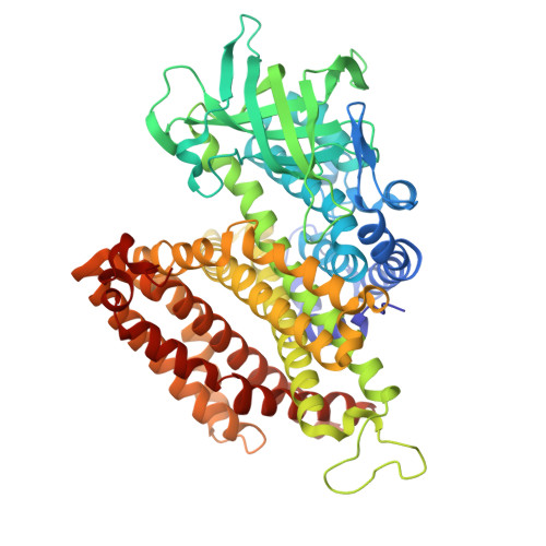
Fatty acids are a primary source of carbon for Pseudomonas aeruginosa (PA) in the airways of people with cystic fibrosis (CF). Here, we use tandem mass-tag proteomics to analyse the protein expression profile of a CF clinical isolate grown on different fatty acids. Two fatty acyl-CoA dehydrogenases (designated FadE1 and FadE2) are strongly induced during growth on fatty acids. FadE1 displays a strong preference for long-chain acyl-CoAs, whereas FadE2 exclusively utilizes medium-chain acyl-CoAs. Structural analysis of the enzymes enables us to identify residues comprising the substrate selectivity filter in each. Engineering these residues enables us to invert the substrate specificity of each enzyme. Mutants in fadE1 displayed impaired virulence in an infection model, and decreased growth on long chain fatty acids. The unique features of the substrate binding pocket enable us to identify an inhibitor that is differentially active against FadE1 and FadE2.
- Department of Biochemistry, Tennis Court Road, Cambridge, UK.
Organizational Affiliation: