8S0N | pdb_00008s0n

Crystal structure of the TMPRSS2 zymogen in complex with the nanobody A07


Experimental Data Snapshot

  • Method: X-RAY DIFFRACTION
  • Resolution: 2.30 Å
  • R-Value Free: 
    0.246 (Depositor), 0.250 (DCC) 
  • R-Value Work: 
    0.211 (Depositor), 0.210 (DCC) 
  • R-Value Observed: 
    0.213 (Depositor) 

Starting Model: experimental
View more details

wwPDB Validation   3D Report Full Report


This is version 1.2 of the entry. See complete history


Literature

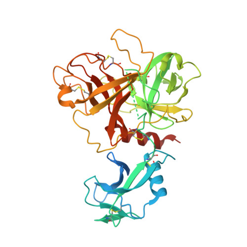
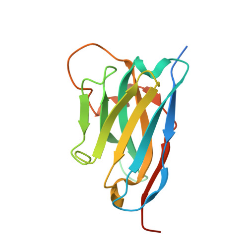
Structural basis of TMPRSS2 zymogen activation and recognition by the HKU1 seasonal coronavirus.

Fernandez, I.Saunders, N.Duquerroy, S.Bolland, W.H.Arbabian, A.Baquero, E.Blanc, C.Lafaye, P.Haouz, A.Buchrieser, J.Schwartz, O.Rey, F.A.

(2024) Cell 187: 4246-4260.e16

  • DOI: https://doi.org/10.1016/j.cell.2024.06.007
  • Primary Citation of Related Structures:  
    8S0L, 8S0M, 8S0N

  • PubMed Abstract: 

    The human seasonal coronavirus HKU1-CoV, which causes common colds worldwide, relies on the sequential binding to surface glycans and transmembrane serine protease 2 (TMPRSS2) for entry into target cells. TMPRSS2 is synthesized as a zymogen that undergoes autolytic activation to process its substrates. Several respiratory viruses, in particular coronaviruses, use TMPRSS2 for proteolytic priming of their surface spike protein to drive membrane fusion upon receptor binding. We describe the crystal structure of the HKU1-CoV receptor binding domain in complex with TMPRSS2, showing that it recognizes residues lining the catalytic groove. Combined mutagenesis of interface residues and comparison across species highlight positions 417 and 469 as determinants of HKU1-CoV host tropism. The structure of a receptor-blocking nanobody in complex with zymogen or activated TMPRSS2 further provides the structural basis of TMPRSS2 activating conformational change, which alters loops recognized by HKU1-CoV and dramatically increases binding affinity.


  • Organizational Affiliation
    • Institut Pasteur, Université de Paris Cité, CNRS UMR 3569, Structural Virology Unit, 75015 Paris, France.

Macromolecules
Find similar proteins by:  (by identity cutoff)  |  3D Structure
Entity ID: 1
MoleculeChains Sequence LengthOrganismDetailsImage
Transmembrane protease serine 2A,
D [auth C]
397Homo sapiensMutation(s): 1 
Gene Names: TMPRSS2PRSS10
EC: 3.4.21.122
UniProt & NIH Common Fund Data Resources
Find proteins for O15393 (Homo sapiens)
Explore O15393 
Go to UniProtKB:  O15393
PHAROS:  O15393
GTEx:  ENSG00000184012 
Entity Groups  
Sequence Clusters30% Identity50% Identity70% Identity90% Identity95% Identity100% Identity
UniProt GroupO15393
Sequence Annotations
Expand
  • Reference Sequence
Find similar proteins by:  (by identity cutoff)  |  3D Structure
Entity ID: 2
MoleculeChains Sequence LengthOrganismDetailsImage
nanobody A07B,
C [auth D]
150Vicugna pacosMutation(s): 0 
Entity Groups  
Sequence Clusters30% Identity50% Identity70% Identity90% Identity95% Identity100% Identity
Sequence Annotations
Expand
  • Reference Sequence
Experimental Data & Validation

Experimental Data

  • Method: X-RAY DIFFRACTION
  • Resolution: 2.30 Å
  • R-Value Free:  0.246 (Depositor), 0.250 (DCC) 
  • R-Value Work:  0.211 (Depositor), 0.210 (DCC) 
  • R-Value Observed: 0.213 (Depositor) 
Space Group: C 1 2 1
Unit Cell:
Length ( Å )Angle ( ˚ )
a = 161.869α = 90
b = 54.458β = 108.393
c = 165.822γ = 90
Software Package:
Software NamePurpose
PHENIXrefinement
XDSdata reduction
Aimlessdata scaling
STARANISOdata scaling
PHASERphasing

Structure Validation

View Full Validation Report


Entry History & Funding Information

Deposition Data


Funding OrganizationLocationGrant Number
Not funded--

Revision History  (Full details and data files)

  • Version 1.0: 2024-06-26
    Type: Initial release
  • Version 1.1: 2024-09-04
    Changes: Database references
  • Version 1.2: 2024-11-06
    Changes: Structure summary