8V2K | pdb_00008v2k

Proteus vulgaris tryptophan indole-lyase complexed with L-alanine

  • Classification: LYASE
  • Organism(s): Proteus vulgaris
  • Expression System: Escherichia coli BL21(DE3)
  • Mutation(s): No 

  • Deposited: 2023-11-22 Released: 2023-12-06 
  • Deposition Author(s): Phillips, R.S.
  • Funding Organization(s): National Institutes of Health/National Institute of General Medical Sciences (NIH/NIGMS)

Experimental Data Snapshot

  • Method: X-RAY DIFFRACTION
  • Resolution: 1.81 Å
  • R-Value Free: 
    0.231 (Depositor), 0.230 (DCC) 
  • R-Value Work: 
    0.186 (Depositor), 0.190 (DCC) 
  • R-Value Observed: 
    0.189 (Depositor) 

Starting Model: experimental
View more details

wwPDB Validation   3D Report Full Report


Ligand Structure Quality Assessment 


This is version 1.1 of the entry. See complete history


Literature

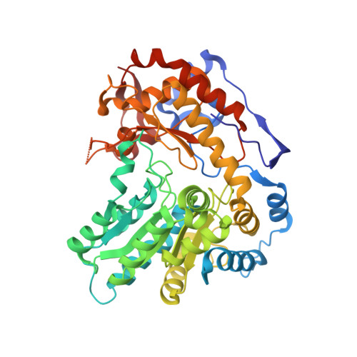
Structure and dynamics of Proteus vulgaris tryptophan indole-lyase complexes with l-ethionine and l-alanine.

Phillips, R.S.Brown, S.M.

(2025) Arch Biochem Biophys 768: 110402-110402

  • DOI: https://doi.org/10.1016/j.abb.2025.110402
  • Primary Citation of Related Structures:  
    8V2K, 8V4A, 9DY7

  • PubMed Abstract: 

    Tryptophan indole-lyase (TIL; [E.C. 4.1.99.1]) is a pyridoxal-5'-phosphate (PLP) dependent enzyme that catalyzes the reversible β-elimination of indole from l-tryptophan. l-Alanine and l-ethionine are TIL competitive inhibitors that form stable quinonoid complexes with λ max ∼508 nm. We have now determined the X-ray crystal structure of the tetrameric TIL complexes with l-alanine and l-ethionine, with either K + or Na + in the cation binding site. For the K + -form, the structures show a mixture of external aldimine and quinonoid complexes, with both open and closed active site conformations. However, the Na + -form exhibits noncovalent and external aldimine complexes in only open active site conformations. Stopped-flow kinetics of l-ethionine binding show that the Na + -form of TIL reacts much more slowly than the K + -form. The l-alanine and l-ethionine complexes of TIL are affected by hydrostatic pressure, suggesting that solvation contributes to the reaction. As pressure increases, the peak at 508 nm decreases, and a new peak at 344 nm appears. These changes are reversible when pressure is released. The 344 nm species could be either a gem-diamine or an enolimine tautomer of the external aldimine. We measured the fluorescence spectrum of the complex under pressure to differentiate these structures. When excited at either 290 or 325 nm, the complex emits at 400 nm, establishing that it is a gem-diamine complex. This peak does not form when the Na + -form of TIL complexed with l-ethionine is subjected to high pressure. Pressure jumps for the TIL-K + -l-ethionine complex measured at 508 nm result in pressure dependent relaxation rate constants. The relaxations show a large activation volume in the direction of quinonoid intermediate formation, suggesting that it is coupled with a conformational change. These results provide new insights into the dynamics of ligand binding to TIL.


  • Organizational Affiliation

    Department of Chemistry, University of Georgia, Athens, GA, 30602, USA; Department of Biochemistry and Molecular Biology, University of Georgia, Athens, GA, 30602, USA. Electronic address: plp@uga.edu.


Macromolecules
Find similar proteins by:  (by identity cutoff)  |  3D Structure
Entity ID: 1
MoleculeChains Sequence LengthOrganismDetailsImage
Tryptophanase
A, B, C, D
467Proteus vulgarisMutation(s): 0 
Gene Names: tnaA
EC: 4.1.99.1
UniProt
Find proteins for P28796 (Proteus vulgaris)
Explore P28796 
Go to UniProtKB:  P28796
Entity Groups  
Sequence Clusters30% Identity50% Identity70% Identity90% Identity95% Identity100% Identity
UniProt GroupP28796
Sequence Annotations
Expand
  • Reference Sequence
Small Molecules
Ligands 4 Unique
IDChains Name / Formula / InChI Key2D Diagram3D Interactions
PLI (Subject of Investigation/LOI)
Query on PLI

Download Ideal Coordinates CCD File 
I [auth B],
M [auth C]
(2E)-2-{[(Z)-{3-HYDROXY-2-METHYL-5-[(PHOSPHONOOXY)METHYL]PYRIDIN-4(1H)-YLIDENE}METHYL]IMINO}PROPANOIC ACID
C11 H15 N2 O7 P
ADVVJYVQWSKPQC-ZAQQZXBOSA-N
F0G (Subject of Investigation/LOI)
Query on F0G

Download Ideal Coordinates CCD File 
F [auth A],
N [auth D]
(E)-N-({3-hydroxy-2-methyl-5-[(phosphonooxy)methyl]pyridin-4-yl}methylidene)-L-alanine
C11 H15 N2 O7 P
UPOKXKNJCNHZTH-ASMSWWRESA-N
DMS
Query on DMS

Download Ideal Coordinates CCD File 
H [auth B],
L [auth C]
DIMETHYL SULFOXIDE
C2 H6 O S
IAZDPXIOMUYVGZ-UHFFFAOYSA-N
K
Query on K

Download Ideal Coordinates CCD File 
E [auth A],
G [auth B],
J [auth C],
K [auth C]
POTASSIUM ION
K
NPYPAHLBTDXSSS-UHFFFAOYSA-N
Experimental Data & Validation

Experimental Data

  • Method: X-RAY DIFFRACTION
  • Resolution: 1.81 Å
  • R-Value Free:  0.231 (Depositor), 0.230 (DCC) 
  • R-Value Work:  0.186 (Depositor), 0.190 (DCC) 
  • R-Value Observed: 0.189 (Depositor) 
Space Group: P 1 21 1
Unit Cell:
Length ( Å )Angle ( ˚ )
a = 66.889α = 90
b = 113.073β = 98.33
c = 120.332γ = 90
Software Package:
Software NamePurpose
PHENIXrefinement
autoPROCdata processing
XDSdata reduction
Aimlessdata scaling
PHENIXphasing

Structure Validation

View Full Validation Report


Ligand Structure Quality Assessment 


Entry History & Funding Information

Deposition Data


Funding OrganizationLocationGrant Number
National Institutes of Health/National Institute of General Medical Sciences (NIH/NIGMS)United StatesR01GM137008

Revision History  (Full details and data files)

  • Version 1.0: 2023-12-06
    Type: Initial release
  • Version 1.1: 2025-04-16
    Changes: Database references, Structure summary