Crystal structure of dihydroneopterin aldolase from Mycobacterium tuberculosis associated with 8-mercaptoguanine, and development of novel S8-functionalized analogues as inhibitors: Synthesis, enzyme inhibition, in vitro toxicity and antitubercular activity.
Czeczot, A.M., Muniz, M.N., Perello, M.A., Silva, E.E.D., Timmers, L.F.S.M., Berger, A., Gonzalez, L.C., Arrache Goncalves, G., Moura, S., Machado, P., Bizarro, C.V., Basso, L.A.(2024) J Enzyme Inhib Med Chem 39: 2388207-2388207
- PubMed: 39140692
- DOI: https://doi.org/10.1080/14756366.2024.2388207
- Primary Citation of Related Structures:
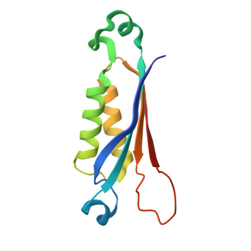
9B2E - PubMed Abstract:
The crystallographic structure of the FolB enzyme from Mycobacterium tuberculosis ( Mt FolB), complexed with its inhibitor 8-mercaptoguanine (8-MG), was elucidated at a resolution of 1.95 Å. A novel series of S8-functionalized 8-MG derivatives were synthesised and evaluated as in vitro inhibitors of dihydroneopterin aldolase (DHNA, EC 4.1.2.25) activity of Mt FolB. These compounds exhibited IC 50 values in the submicromolar range. Evaluation of the activity for five compounds indicated their inhibition mode and inhibition constants. Molecular docking analyses were performed to determine the enzyme-inhibitor intermolecular interactions and ligand conformations upon complex formation. The inhibitory activities of all compounds against the M. tuberculosis H37Rv strain were evaluated. Compound 3e exhibited a minimum inhibitory concentration in the micromolar range. Finally, Compound 3e showed no apparent toxicity in both HepG2 and Vero cells. The findings presented herein will advance the quest for novel, specific inhibitors targeting Mt FolB, an attractive molecular target for TB drug development.
- Centro de Pesquisas em Biologia Molecular e Funcional, Instituto Nacional de Ciência e Tecnologia em Tuberculose, Escola de Ciências da Saúde e da Vida, Pontifícia Universidade Católica do Rio Grande do Sul, Porto Alegre, Brazil.
Organizational Affiliation: