9DRS | pdb_00009drs

Crystal structure of M. tuberculosis PheRS-tRNA complex bound to inhibitor D-116


Experimental Data Snapshot

  • Method: X-RAY DIFFRACTION
  • Resolution: 2.35 Å
  • R-Value Free: 
    0.250 (Depositor), 0.250 (DCC) 
  • R-Value Work: 
    0.201 (Depositor), 0.201 (DCC) 
  • R-Value Observed: 
    0.203 (Depositor) 

Starting Model: experimental
View more details

wwPDB Validation   3D Report Full Report


Ligand Structure Quality Assessment 


This is version 1.1 of the entry. See complete history


Literature

Different chemical scaffolds bind to L-phe site in Mycobacterium tuberculosis Phe-tRNA synthetase.

Gade, P.Chang, C.Pryde, D.S.Fletcher, D.Niven, S.Magalhaes, L.G.Robinson, D.Saini, J.Ibrahim, P.E.G.F.Forte, B.Wower, J.Bodkin, M.J.Baragana, B.Gilbert, I.H.Michalska, K.Joachimiak, A.

(2025) Eur J Med Chem 287: 117335-117335

  • DOI: https://doi.org/10.1016/j.ejmech.2025.117335
  • Primary Citation of Related Structures:  
    9DRS, 9DRV

  • PubMed Abstract: 

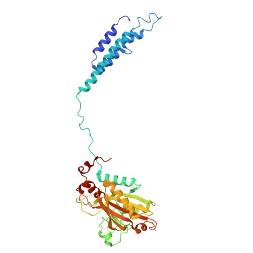
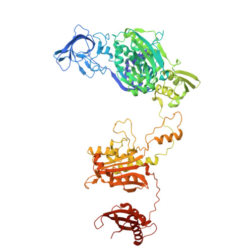
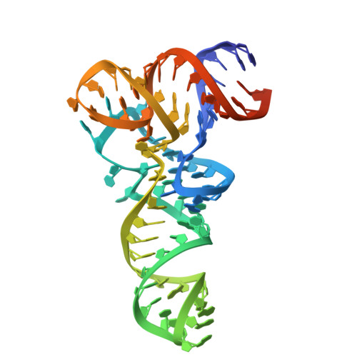
    Tuberculosis (TB), caused by Mycobacterium tuberculosis (Mt), is one of the deadliest infectious diseases. The rise of multidrug-resistant strains represents a major public health threat, requiring new therapeutic options. Bacterial aminoacyl-tRNA synthetases (aaRS) have been shown to be highly promising drug targets, including for TB treatment. These enzymes play an essential role in translating the DNA gene code into protein sequence by attaching specific amino acid to their cognate tRNAs. They have multiple binding sites that can be targeted for inhibitor discovery: amino acid binding pocket, ATP binding pocket, tRNA binding site and an editing domain. Recently we reported several high-resolution structures of M. tuberculosis phenylalanyl-tRNA synthetase (MtPheRS) complexed with tRNA Phe and either L-Phe or a nonhydrolyzable phenylalanine adenylate analog. Here, using Nucleic Magnetic Resonance (NMR) and Surface Plasmon Resonance (SPR) we identified fragments that bind to MtPheRS and we determined crystal structures of their complexes with MtPheRS/tRNA Phe . All the binders interact with the L-Phe amino acid binding site. The analysis of interactions of the new compounds combined with adenylate analog structure provides insights for the rational design of anti-tuberculosis drugs. The 3' arm of the tRNA Phe in all the structures was disordered with exception of one complex with D-735 compound. In this structure the 3' CCA end of the acceptor stem is observed in the editing domain of MtPheRS providing insights regarding the post-transfer editing activity of class II aaRS.


  • Organizational Affiliation

    Center for Structural Biology of Infectious Diseases, Consortium for Advanced Science and Engineering, University of Chicago, Chicago, IL, 60667, USA; Structural Biology Center, X-ray Science Division, Argonne National Laboratory, Lemont, IL, 60439, USA.


Macromolecules

Find similar proteins by:  (by identity cutoff)  |  3D Structure
Entity ID: 1
MoleculeChains Sequence LengthOrganismDetailsImage
Phenylalanine--tRNA ligase alpha subunit
A, D
341Mycobacterium tuberculosis H37RvMutation(s): 0 
Gene Names: pheSRv1649MTCY06H11.14
EC: 6.1.1.20
UniProt
Find proteins for P9WFU3 (Mycobacterium tuberculosis (strain ATCC 25618 / H37Rv))
Explore P9WFU3 
Go to UniProtKB:  P9WFU3
Entity Groups  
Sequence Clusters30% Identity50% Identity70% Identity90% Identity95% Identity100% Identity
UniProt GroupP9WFU3
Sequence Annotations
Expand
  • Reference Sequence
Find similar proteins by:  (by identity cutoff)  |  3D Structure
Entity ID: 2
MoleculeChains Sequence LengthOrganismDetailsImage
Phenylalanine--tRNA ligase beta subunit
B, E
835Mycobacterium tuberculosis H37RvMutation(s): 0 
Gene Names: pheTRv1650MTCY06H11.15
EC: 6.1.1.20
UniProt
Find proteins for P9WFU1 (Mycobacterium tuberculosis (strain ATCC 25618 / H37Rv))
Explore P9WFU1 
Go to UniProtKB:  P9WFU1
Entity Groups  
Sequence Clusters30% Identity50% Identity70% Identity90% Identity95% Identity100% Identity
UniProt GroupP9WFU1
Sequence Annotations
Expand
  • Reference Sequence
Find similar nucleic acids by:  (by identity cutoff)  |  3D Structure
Entity ID: 3
MoleculeChains LengthOrganismImage
tRNA(Phe)
C, F
77Mycobacterium tuberculosis H37Rv
Sequence Annotations
Expand
  • Reference Sequence
Small Molecules
Ligands 7 Unique
IDChains Name / Formula / InChI Key2D Diagram3D Interactions
EPE
Query on EPE

Download Ideal Coordinates CCD File 
Y [auth B]4-(2-HYDROXYETHYL)-1-PIPERAZINE ETHANESULFONIC ACID
C8 H18 N2 O4 S
JKMHFZQWWAIEOD-UHFFFAOYSA-N
AX7 (Subject of Investigation/LOI)
Query on AX7

Download Ideal Coordinates CCD File 
G [auth A],
GA [auth D]
1H-benzimidazol-2-amine
C7 H7 N3
JWYUFVNJZUSCSM-UHFFFAOYSA-N
PEG
Query on PEG

Download Ideal Coordinates CCD File 
LA [auth E],
O [auth B],
P [auth B],
Z [auth B]
DI(HYDROXYETHYL)ETHER
C4 H10 O3
MTHSVFCYNBDYFN-UHFFFAOYSA-N
DMS
Query on DMS

Download Ideal Coordinates CCD File 
JA [auth D]DIMETHYL SULFOXIDE
C2 H6 O S
IAZDPXIOMUYVGZ-UHFFFAOYSA-N
EDO
Query on EDO

Download Ideal Coordinates CCD File 
AA [auth C]
KA [auth E]
L [auth B]
PA [auth E]
QA [auth E]
AA [auth C],
KA [auth E],
L [auth B],
PA [auth E],
QA [auth E],
RA [auth E],
W [auth B],
X [auth B]
1,2-ETHANEDIOL
C2 H6 O2
LYCAIKOWRPUZTN-UHFFFAOYSA-N
ACT
Query on ACT

Download Ideal Coordinates CCD File 
BA [auth C]
I [auth A]
IA [auth D]
J [auth A]
K [auth B]
BA [auth C],
I [auth A],
IA [auth D],
J [auth A],
K [auth B],
M [auth B],
MA [auth E],
N [auth B],
NA [auth E],
OA [auth E],
Q [auth B],
R [auth B],
S [auth B],
T [auth B],
U [auth B],
V [auth B]
ACETATE ION
C2 H3 O2
QTBSBXVTEAMEQO-UHFFFAOYSA-M
MG
Query on MG

Download Ideal Coordinates CCD File 
CA [auth C]
DA [auth C]
EA [auth C]
FA [auth C]
H [auth A]
CA [auth C],
DA [auth C],
EA [auth C],
FA [auth C],
H [auth A],
HA [auth D],
SA [auth F]
MAGNESIUM ION
Mg
JLVVSXFLKOJNIY-UHFFFAOYSA-N
Experimental Data & Validation

Experimental Data

  • Method: X-RAY DIFFRACTION
  • Resolution: 2.35 Å
  • R-Value Free:  0.250 (Depositor), 0.250 (DCC) 
  • R-Value Work:  0.201 (Depositor), 0.201 (DCC) 
  • R-Value Observed: 0.203 (Depositor) 
Space Group: P 1 21 1
Unit Cell:
Length ( Å )Angle ( ˚ )
a = 146.676α = 90
b = 64.106β = 111.21
c = 188.334γ = 90
Software Package:
Software NamePurpose
PHENIXrefinement
HKL-3000data reduction
HKL-3000data scaling
MOLREPphasing

Structure Validation

View Full Validation Report


Ligand Structure Quality Assessment 


Entry History & Funding Information

Deposition Data


Funding OrganizationLocationGrant Number
National Institutes of Health/National Institute Of Allergy and Infectious Diseases (NIH/NIAID)United States--

Revision History  (Full details and data files)

  • Version 1.0: 2025-02-12
    Type: Initial release
  • Version 1.1: 2025-02-19
    Changes: Database references