


Structure of ALAS bound to glycine from S. cerevisiae
Tran, J.U., Brown, B.L.To be published.
Experimental Data Snapshot
Starting Model: experimental
View more details
Entity ID: 1 | |||||
|---|---|---|---|---|---|
| Molecule | Chains | Sequence Length | Organism | Details | Image |
| 5-aminolevulinate synthase, mitochondrial | 491 | Saccharomyces cerevisiae | Mutation(s): 0 Gene Names: HEM1, CYD1, YDR232W, YD9934.16 EC: 2.3.1.37 | 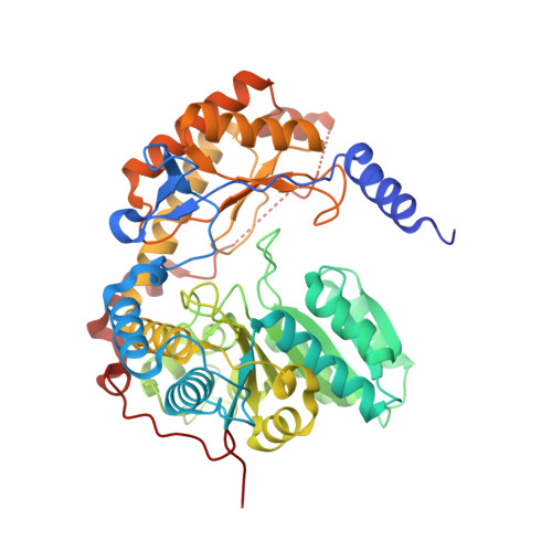 | |
UniProt | |||||
Find proteins for P09950 (Saccharomyces cerevisiae (strain ATCC 204508 / S288c)) Explore P09950 Go to UniProtKB: P09950 | |||||
Entity Groups | |||||
| Sequence Clusters | 30% Identity50% Identity70% Identity90% Identity95% Identity100% Identity | ||||
| UniProt Group | P09950 | ||||
Sequence AnnotationsExpand | |||||
| |||||
| Ligands 2 Unique | |||||
|---|---|---|---|---|---|
| ID | Chains | Name / Formula / InChI Key | 2D Diagram | 3D Interactions | |
| PLG (Subject of Investigation/LOI) Query on PLG | FA [auth D] G [auth F] H [auth A] HA [auth B] O [auth C] | N-GLYCINE-[3-HYDROXY-2-METHYL-5-PHOSPHONOOXYMETHYL-PYRIDIN-4-YL-METHANE] C10 H15 N2 O7 P FEVQWBMNLWUBTF-UHFFFAOYSA-N |  | ||
| PEG Query on PEG | AA [auth E] BA [auth E] CA [auth E] DA [auth E] EA [auth E] | DI(HYDROXYETHYL)ETHER C4 H10 O3 MTHSVFCYNBDYFN-UHFFFAOYSA-N |  | ||
| Length ( Å ) | Angle ( ˚ ) |
|---|---|
| a = 59.672 | α = 115.76 |
| b = 112.518 | β = 98.31 |
| c = 117.388 | γ = 91.49 |
| Software Name | Purpose |
|---|---|
| REFMAC | refinement |
| XDS | data reduction |
| XDS | data scaling |
| PHASER | phasing |
| Funding Organization | Location | Grant Number |
|---|---|---|
| National Institutes of Health/National Institute of General Medical Sciences (NIH/NIGMS) | United States | DP2GM146255 |
| American Heart Association | United States | 24PRE1190012 |


