9EMZ | pdb_00009emz

13C/15N-labelled Integral Membrane Enzyme LspA in the Lipid Cubic Phase


Experimental Data Snapshot

  • Method: X-RAY DIFFRACTION
  • Resolution: 3.00 Å
  • R-Value Free: 
    0.303 (Depositor), 0.303 (DCC) 
  • R-Value Work: 
    0.245 (Depositor), 0.247 (DCC) 
  • R-Value Observed: 
    0.248 (Depositor) 

Starting Model: experimental
View more details

wwPDB Validation   3D Report Full Report


Ligand Structure Quality Assessment 


This is version 1.2 of the entry. See complete history


Literature

Solid-state NMR of membrane peptides and proteins in the lipid cubic phase.

Ramberg, K.O.Boland, C.Kooshapur, H.Soubias, O.Wiktor, M.Huang, C.Y.Bailey, J.Gawrisch, K.Caffrey, M.

(2025) Biophys J 124: 1387-1400

  • DOI: https://doi.org/10.1016/j.bpj.2025.03.012
  • Primary Citation of Related Structures:  
    9EMZ

  • PubMed Abstract: 

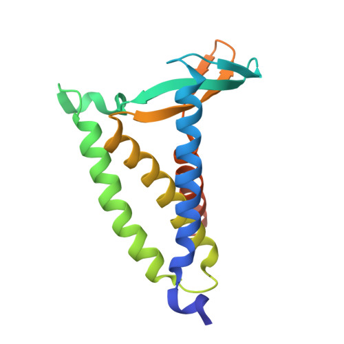
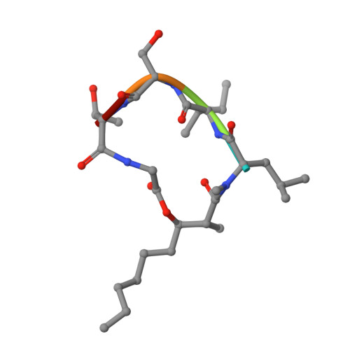
    Solid-state nuclear magnetic resonance (ssNMR) is a powerful technique for studying membrane protein structure and dynamics. Ideally, measurements are performed with the protein in a lipid bilayer. However, homogenous reconstitution of functional protein into intact bilayers at sufficiently high concentrations is often difficult to achieve. In this work, we investigate the suitability of the lipid cubic phase (LCP), which incorporates a lipid bilayer, as an alternative medium for ssNMR of integral membrane peptides and proteins. The cubic mesophase has long been used to generate membrane protein crystals for use in X-ray crystallographic structure determination by the so-called in meso method, and for protein functional and biophysical characterization. Preparing and handling protein-laden LCP is straightforward. LCP may therefore provide a valuable alternative to native membranes and other membrane mimetics for ssNMR. We tested this idea by conducting standard magic angle spinning (MAS) ssNMR experiments on LCP into which gramicidin, a ∼4 kDa transmembrane peptide, or bacterial lipoprotein signal peptidase II (LspA), a ∼20 kDa integral membrane enzyme, had been reconstituted. We report one- and two-dimensional ssNMR spectra for both gramicidin and LspA and the parameters for optimizing spectral quality. The high protein carrying capacity of the cubic phase facilitated 13 C ssNMR at natural abundance. Lowering temperature and raising MAS frequency enabled significant improvements in spectral quality. One-dimensional 13 C and 15 N spectra were collected for LspA. Two-dimensional ssNMR experiments provided information on LspA dynamics and its interaction with the water and lipid components of the cubic phase. Solution NMR measurements carried out in parallel yielded information on the effect of the antibiotic, globomycin, on LspA structure and dynamics.


  • Organizational Affiliation

    Membrane Structural & Functional Biology Group, School of Medicine and School of Biochemistry & Immunology, Trinity College Dublin, Dublin, D02 R590, Ireland. Electronic address: martin.caffrey@tcd.ie.


Macromolecules
Find similar proteins by:  (by identity cutoff)  |  3D Structure
Entity ID: 1
MoleculeChains Sequence LengthOrganismDetailsImage
Lipoprotein signal peptidase
A, B, C, D
169Pseudomonas aeruginosa PAO1Mutation(s): 0 
Gene Names: lspAlsPA4559
EC: 3.4.23.36
Membrane Entity: Yes 
UniProt
Find proteins for Q9HVM5 (Pseudomonas aeruginosa (strain ATCC 15692 / DSM 22644 / CIP 104116 / JCM 14847 / LMG 12228 / 1C / PRS 101 / PAO1))
Explore Q9HVM5 
Go to UniProtKB:  Q9HVM5
Entity Groups  
Sequence Clusters30% Identity50% Identity70% Identity90% Identity95% Identity100% Identity
UniProt GroupQ9HVM5
Sequence Annotations
Expand
  • Reference Sequence

Find similar proteins by:  Sequence   |   3D Structure  

Entity ID: 2
MoleculeChains Sequence LengthOrganismDetailsImage
Globomycin
E, F, G, H
5synthetic constructMutation(s): 0 
Sequence Annotations
Expand
  • Reference Sequence
Small Molecules
Ligands 1 Unique
IDChains Name / Formula / InChI Key2D Diagram3D Interactions
OLC
Query on OLC

Download Ideal Coordinates CCD File 
AA [auth C]
BA [auth D]
CA [auth D]
DA [auth D]
EA [auth D]
AA [auth C],
BA [auth D],
CA [auth D],
DA [auth D],
EA [auth D],
FA [auth D],
I [auth A],
J [auth A],
K [auth A],
L [auth A],
M [auth A],
N [auth A],
O [auth B],
P [auth B],
Q [auth B],
R [auth B],
S [auth B],
T [auth B],
U [auth B],
V [auth C],
W [auth C],
X [auth C],
Y [auth C],
Z [auth C]
(2R)-2,3-dihydroxypropyl (9Z)-octadec-9-enoate
C21 H40 O4
RZRNAYUHWVFMIP-GDCKJWNLSA-N
Modified Residues  3 Unique
IDChains TypeFormula2D DiagramParent
ALO
Query on ALO
E, F, G, H
L-PEPTIDE LINKINGC4 H9 N O3THR
IIL
Query on IIL
E, F, G, H
L-PEPTIDE LINKINGC6 H13 N O2ILE
MLE
Query on MLE
E, F, G, H
L-PEPTIDE LINKINGC7 H15 N O2LEU
Experimental Data & Validation

Experimental Data

  • Method: X-RAY DIFFRACTION
  • Resolution: 3.00 Å
  • R-Value Free:  0.303 (Depositor), 0.303 (DCC) 
  • R-Value Work:  0.245 (Depositor), 0.247 (DCC) 
  • R-Value Observed: 0.248 (Depositor) 
Space Group: C 1 2 1
Unit Cell:
Length ( Å )Angle ( ˚ )
a = 113.991α = 90
b = 106.086β = 97.54
c = 85.919γ = 90
Software Package:
Software NamePurpose
PHENIXrefinement
XDSdata reduction
XSCALEdata scaling
PHASERphasing

Structure Validation

View Full Validation Report


Ligand Structure Quality Assessment 


Entry History & Funding Information

Deposition Data


Funding OrganizationLocationGrant Number
Science Foundation IrelandIreland16/IA/4435 and 22/FFP-A/10278
Swiss National Science FoundationSwitzerlandP2BSP3_15254

Revision History  (Full details and data files)

  • Version 1.0: 2025-03-26
    Type: Initial release
  • Version 1.1: 2025-04-02
    Changes: Database references
  • Version 1.2: 2025-05-21
    Changes: Database references