Crystal structure of a bacterial photoactivated adenylate cyclase determined by serial femtosecond and serial synchrotron crystallography.
Kapetanaki, S.M., Coquelle, N., von Stetten, D., Byrdin, M., Rios-Santacruz, R., Bean, R., Bielecki, J., Boudjelida, M., Fekete, Z., Grime, G.W., Han, H., Hatton, C., Kantamneni, S., Kharitonov, K., Kim, C., Kloos, M., Koua, F.H.M., de Diego Martinez, I., Melo, D., Rane, L., Round, A., Round, E., Sarma, A., Schubert, R., Schulz, J., Sikorski, M., Vakili, M., Valerio, J., Vitas, J., de Wijn, R., Wrona, A., Zala, N., Pearson, A., Dorner, K., Schiro, G., Garman, E.F., Lukacs, A., Weik, M.(2024) IUCrJ 11: 991-1006
- PubMed: 39470573
- DOI: https://doi.org/10.1107/S2052252524010170
- Primary Citation of Related Structures:
9F1Z - PubMed Abstract:
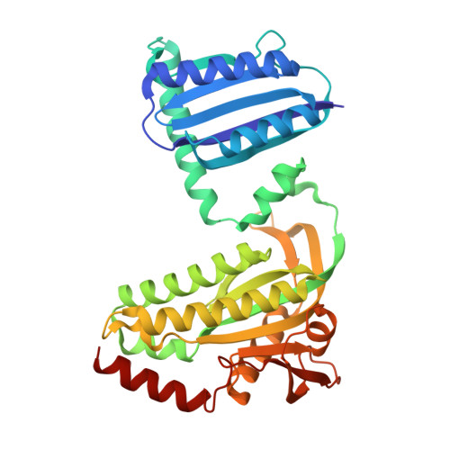
OaPAC is a recently discovered blue-light-using flavin adenosine dinucleotide (BLUF) photoactivated adenylate cyclase from the cyanobacterium Oscillatoria acuminata that uses adenosine triphosphate and translates the light signal into the production of cyclic adenosine monophosphate. Here, we report crystal structures of the enzyme in the absence of its natural substrate determined from room-temperature serial crystallography data collected at both an X-ray free-electron laser and a synchrotron, and we compare these structures with cryo-macromolecular crystallography structures obtained at a synchrotron by us and others. These results reveal slight differences in the structure of the enzyme due to data collection at different temperatures and X-ray sources. We further investigate the effect of the Y6W mutation in the BLUF domain, a mutation which results in a rearrangement of the hydrogen-bond network around the flavin and a notable rotation of the side chain of the critical Gln48 residue. These studies pave the way for picosecond-millisecond time-resolved serial crystallography experiments at X-ray free-electron lasers and synchrotrons in order to determine the early structural intermediates and correlate them with the well studied picosecond-millisecond spectroscopic intermediates.
- Université Grenoble Alpes, CEA, CNRS, Institut de Biologie Structurale, 38044 Grenoble, France.
Organizational Affiliation: