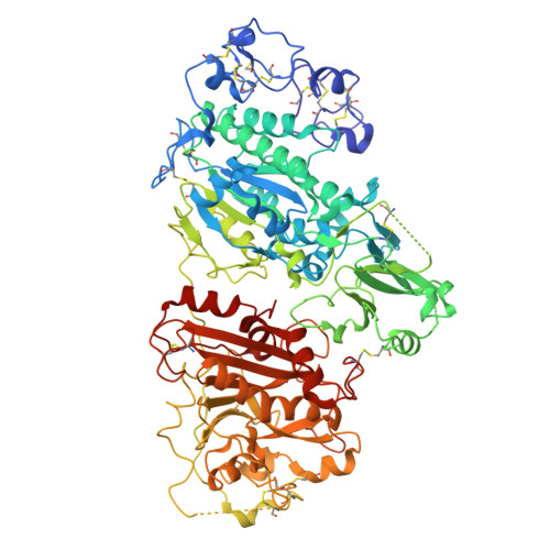
9FTN | pdb_00009ftn

Crystal Structure of Autotaxin (ENPP2) with Type VI Inhibitor, a Novel Class of Inhibitors with Three-Point Lock Binding Mode


Experimental Data Snapshot

  • Method: X-RAY DIFFRACTION
  • Resolution: 2.90 Å
  • R-Value Free: 
    0.268 (Depositor), 0.272 (DCC) 
  • R-Value Work: 
    0.207 (Depositor), 0.211 (DCC) 

Starting Model: experimental
View more details

wwPDB Validation   3D Report Full Report


Ligand Structure Quality Assessment 


This is version 1.1 of the entry. See complete history


Literature

Design, Synthesis, and Biological Implications of Autotaxin inhibitors with a Three-Point lock binding mode.

Desroy, N.Borza, R.Heiermann, J.Triballeau, N.Joncour, A.Bienvenu, N.Hengeveld, W.J.Springer, J.Galien, R.Joosten, R.P.Perrakis, A.Heckmann, B.

(2025) Bioorg Med Chem 124: 118181-118181

  • DOI: https://doi.org/10.1016/j.bmc.2025.118181
  • Primary Citation of Related Structures:  
    9FTN, 9FXU, 9FXW, 9FXY

  • PubMed Abstract: 

    Autotaxin (ATX) is a circulating enzyme that plays a major role in the production of the signaling mediator lysophosphatidic acid (LPA). A role for ATX/LPA signaling has been described in multiple disease areas, including fibrosis and cancer. ATX inhibitors are classified in five types (I-V) depending on how they target parts of the tripartite site (active site, pocket and tunnel). We set to explore a "penultimate" type of inhibitors, targeting all these three parts at once. Designing new analogs extending on an ethyl group of the type IV GLPG1690 compound, yielded potent new molecules. Co-crystal structures confirmed compounds that utilize a three-point lock binding mode. The most potent "type VI" inhibitors, 4 and 41, displayed increased inhibitory activity (∼40-fold) compared to the type IV close analog 3. Type VI inhibitors 4 and 41 showed cellular and phenotypic activity similar to type IV inhibitor GLPG1690. Identification of this new binding mode completes this combinatorial puzzle in inhibitor design and calls for further investigation to characterize potential therapeutic benefit.


  • Organizational Affiliation
    • Galapagos SASU, 102 Avenue Gaston Roussel, 93230 Romainville, France. Electronic address: nicolas.desroy@glpg.com.

Macromolecules
Find similar proteins by:  (by identity cutoff)  |  3D Structure
Entity ID: 1
MoleculeChains Sequence LengthOrganismDetailsImage
Isoform 2 of Ectonucleotide pyrophosphatase/phosphodiesterase family member 2807Rattus norvegicusMutation(s): 2 
Gene Names: Enpp2AtxNpps2
EC: 3.1.4.39 (PDB Primary Data), 3.1.4.4 (UniProt)
UniProt
Find proteins for Q64610 (Rattus norvegicus)
Explore Q64610 
Go to UniProtKB:  Q64610
Entity Groups  
Sequence Clusters30% Identity50% Identity70% Identity90% Identity95% Identity100% Identity
UniProt GroupQ64610
Glycosylation
Glycosylation Sites: 1Go to GlyGen: Q64610-1
Sequence Annotations
Expand
  • Reference Sequence
Oligosaccharides

Help

Entity ID: 2
MoleculeChains Length2D Diagram Glycosylation3D Interactions
alpha-D-mannopyranose-(1-3)-[alpha-D-mannopyranose-(1-6)]beta-D-mannopyranose-(1-4)-2-acetamido-2-deoxy-beta-D-glucopyranose-(1-4)-2-acetamido-2-deoxy-beta-D-glucopyranose
B
5N-Glycosylation
Glycosylation Resources
GlyTouCan:  G22768VO
GlyCosmos:  G22768VO
GlyGen:  G22768VO
Small Molecules
Ligands 5 Unique
IDChains Name / Formula / InChI Key2D Diagram3D Interactions
A1IG5 (Subject of Investigation/LOI)
Query on A1IG5

Download Ideal Coordinates CCD File 
G [auth A]3-(3-((4-(4-fluorophenyl)thiazol-2-yl)(methyl)amino)-6-(1-(methylsulfonyl)piperidin-4-yl)imidazo[1,2-b]pyridazin-2-yl)-N-(2-oxo-2,3-dihydrobenzo[d]oxazol-6-yl)propenamide
C32 H31 F N8 O5 S2
MVJGOHXOHATEJY-UHFFFAOYSA-N
IOD
Query on IOD

Download Ideal Coordinates CCD File 
F [auth A]IODIDE ION
I
XMBWDFGMSWQBCA-UHFFFAOYSA-M
ZN
Query on ZN

Download Ideal Coordinates CCD File 
C [auth A],
D [auth A]
ZINC ION
Zn
PTFCDOFLOPIGGS-UHFFFAOYSA-N
SCN
Query on SCN

Download Ideal Coordinates CCD File 
H [auth A]
I [auth A]
J [auth A]
K [auth A]
L [auth A]
H [auth A],
I [auth A],
J [auth A],
K [auth A],
L [auth A],
M [auth A],
N [auth A],
O [auth A]
THIOCYANATE ION
C N S
ZMZDMBWJUHKJPS-UHFFFAOYSA-M
CA
Query on CA

Download Ideal Coordinates CCD File 
E [auth A]CALCIUM ION
Ca
BHPQYMZQTOCNFJ-UHFFFAOYSA-N
Experimental Data & Validation

Experimental Data

  • Method: X-RAY DIFFRACTION
  • Resolution: 2.90 Å
  • R-Value Free:  0.268 (Depositor), 0.272 (DCC) 
  • R-Value Work:  0.207 (Depositor), 0.211 (DCC) 
Space Group: P 1
Unit Cell:
Length ( Å )Angle ( ˚ )
a = 53.935α = 98.991
b = 63.558β = 105.207
c = 70.599γ = 99.647
Software Package:
Software NamePurpose
REFMACrefinement
Aimlessdata scaling
MOLREPphasing
Cootmodel building
MolProbitymodel building
XDSdata reduction
PDB-REDOrefinement

Structure Validation

View Full Validation Report


Ligand Structure Quality Assessment 


Entry History & Funding Information

Deposition Data


Funding OrganizationLocationGrant Number
Oncode InstituteNetherlands--

Revision History  (Full details and data files)

  • Version 1.0: 2025-04-23
    Type: Initial release
  • Version 1.1: 2025-04-30
    Changes: Database references