9GR6 | pdb_00009gr6

PsiM in complex with SAH, high resolution


Experimental Data Snapshot

  • Method: X-RAY DIFFRACTION
  • Resolution: 0.93 Å
  • R-Value Free: 
    0.160 (Depositor), 0.161 (DCC) 
  • R-Value Work: 
    0.146 (Depositor), 0.144 (DCC) 
  • R-Value Observed: 
    0.146 (Depositor) 

Starting Model: experimental
View more details

wwPDB Validation   3D Report Full Report


Ligand Structure Quality Assessment 


This is version 1.2 of the entry. See complete history


Literature

If you cannot see it, is it still there?

Leitner, L.Hudspeth, J.Werten, S.Rupp, B.

(2025) J Appl Crystallogr 58: 615-621

  • DOI: https://doi.org/10.1107/S160057672500130X
  • Primary Citation of Related Structures:  
    9GR6, 9GR7

  • PubMed Abstract: 

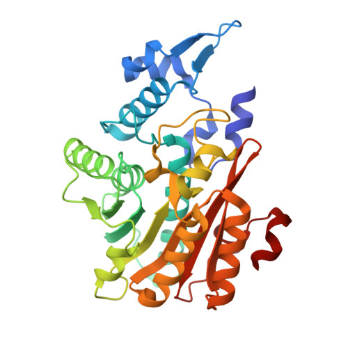
    Protein crystallographers rely on electron density to build atomic models of molecular structures, yet flexible regions often remain unseen in electron density and are omitted. We suggest that ensemble refinement can be used to visualize and analyse the conformational landscape of such 'invisible' protein segments, which is particularly useful in cases where molecular flexibility plays a functional role. Using ensemble refinement on multiple crystal forms of the fungal methyl-transferase PsiM as an example, we illustrate the dynamic nature of a key substrate recognition loop, demonstrating its potential role in substrate binding and release. Ensemble refinement provides a persuasive visualization of biologically relevant flexible regions and can be a powerful tool for exploring molecular plasticity and aiding the modelling of dynamic protein components.


  • Organizational Affiliation

    Institute of Genetic Epidemiology Medical University of Innsbruck Schöpfstrasse 41 6020Innsbruck Austria.


Macromolecules
Find similar proteins by:  (by identity cutoff)  |  3D Structure
Entity ID: 1
MoleculeChains Sequence LengthOrganismDetailsImage
Psilocybin synthase322Psilocybe cubensisMutation(s): 0 
Gene Names: psiM
EC: 2.1.1 (PDB Primary Data), 2.1.1.345 (UniProt)
UniProt
Find proteins for P0DPA9 (Psilocybe cubensis)
Explore P0DPA9 
Go to UniProtKB:  P0DPA9
Entity Groups  
Sequence Clusters30% Identity50% Identity70% Identity90% Identity95% Identity100% Identity
UniProt GroupP0DPA9
Sequence Annotations
Expand
  • Reference Sequence
Experimental Data & Validation

Experimental Data

  • Method: X-RAY DIFFRACTION
  • Resolution: 0.93 Å
  • R-Value Free:  0.160 (Depositor), 0.161 (DCC) 
  • R-Value Work:  0.146 (Depositor), 0.144 (DCC) 
  • R-Value Observed: 0.146 (Depositor) 
Space Group: P 21 21 21
Unit Cell:
Length ( Å )Angle ( ˚ )
a = 49.895α = 90
b = 78.575β = 90
c = 84.013γ = 90
Software Package:
Software NamePurpose
PHENIXrefinement
PHENIXphasing
xia2data reduction
DIALSdata scaling

Structure Validation

View Full Validation Report


Ligand Structure Quality Assessment 


Entry History & Funding Information

Deposition Data


Funding OrganizationLocationGrant Number
Austrian Science FundAustriaI 5192
German Research Foundation (DFG)Germany456463463

Revision History  (Full details and data files)

  • Version 1.0: 2024-12-25
    Type: Initial release
  • Version 1.1: 2025-03-12
    Changes: Database references
  • Version 1.2: 2025-04-16
    Changes: Database references