9GRO | pdb_00009gro

Crystal structure of the engineered C-terminal phosphatase domain from Saccharomyces cerevisiae Vip1 in complex with 1,5-InsP8 (phosphatase dead mutant, loop deletion residues 848-918)


Experimental Data Snapshot

  • Method: X-RAY DIFFRACTION
  • Resolution: 2.36 Å
  • R-Value Free: 
    0.275 (Depositor), 0.267 (DCC) 
  • R-Value Work: 
    0.228 (Depositor), 0.223 (DCC) 
  • R-Value Observed: 
    0.230 (Depositor) 

Starting Model: experimental
View more details

wwPDB Validation   3D Report Full Report


Ligand Structure Quality Assessment 


This is version 1.0 of the entry. See complete history


Literature

A small signaling domain controls PPIP5K phosphatase activity in phosphate homeostasis.

Raia, P.Lee, K.Bartsch, S.M.Rico-Resendiz, F.Portugal-Calisto, D.Vadas, O.Panse, V.G.Fiedler, D.Hothorn, M.

(2025) Nat Commun 16: 1753-1753

  • DOI: https://doi.org/10.1038/s41467-025-56937-0
  • Primary Citation of Related Structures:  
    9GR8, 9GRH, 9GRN, 9GRO

  • PubMed Abstract: 

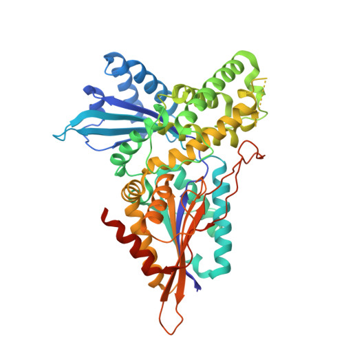
    Inositol pyrophosphates (PP-InsPs) are eukaryotic nutrient messengers. The N-terminal kinase domain of diphosphoinositol pentakisphosphate kinase (PPIP5K) generates the messenger 1,5-InsP 8 , the C-terminal phosphatase domain catalyzes PP-InsP breakdown. The balance between kinase and phosphatase activities regulates 1,5-InsP 8 levels. Here, we present crystal structures of the apo and substrate-bound PPIP5K phosphatase domain from S. cerevisiae (ScVip1 PD ). ScVip1 PD is a phytase-like inositol 1-pyrophosphate histidine phosphatase with two conserved catalytic motifs. The enzyme has a strong preference for 1,5-InsP 8 and is inhibited by inorganic phosphate. It contains an α-helical insertion domain stabilized by a structural Zn 2+ binding site, and a unique GAF domain that channels the substrate to the active site. Mutations that alter the active site, restrict the movement of the GAF domain, or change the substrate channel's charge inhibit the enzyme activity in vitro, and Arabidopsis VIH2 in planta. Our work reveals the structure, enzymatic mechanism and regulation of eukaryotic PPIP5K phosphatases.


  • Organizational Affiliation

    Structural Plant Biology Laboratory, Department of Plant Sciences, University of Geneva, Geneva, Switzerland.


Macromolecules
Find similar proteins by:  (by identity cutoff)  |  3D Structure
Entity ID: 1
MoleculeChains Sequence LengthOrganismDetailsImage
Inositol hexakisphosphate and diphosphoinositol-pentakisphosphate kinase
A, B
507Saccharomyces cerevisiaeMutation(s): 0 
Gene Names: VIP1YLR410W
EC: 2.7.4.24
UniProt
Find proteins for Q06685 (Saccharomyces cerevisiae (strain ATCC 204508 / S288c))
Explore Q06685 
Go to UniProtKB:  Q06685
Entity Groups  
Sequence Clusters30% Identity50% Identity70% Identity90% Identity95% Identity100% Identity
UniProt GroupQ06685
Sequence Annotations
Expand
  • Reference Sequence
Small Molecules
Ligands 7 Unique
IDChains Name / Formula / InChI Key2D Diagram3D Interactions
I8P
Query on I8P

Download Ideal Coordinates CCD File 
DA [auth B],
Q [auth A]
(1R,3S,4R,5S,6R)-2,4,5,6-tetrakis(phosphonooxy)cyclohexane-1,3-diyl bis[trihydrogen (diphosphate)]
C6 H20 O30 P8
HHQOOERQSFJGEP-SLWYWOEDSA-N
SPM
Query on SPM

Download Ideal Coordinates CCD File 
D [auth A],
E [auth A]
SPERMINE
C10 H26 N4
PFNFFQXMRSDOHW-UHFFFAOYSA-N
1JW
Query on 1JW

Download Ideal Coordinates CCD File 
V [auth B],
X [auth B]
(2S)-hexane-1,2,6-triol
C6 H14 O3
ZWVMLYRJXORSEP-LURJTMIESA-N
ORN
Query on ORN

Download Ideal Coordinates CCD File 
F [auth A]L-ornithine
C5 H12 N2 O2
AHLPHDHHMVZTML-BYPYZUCNSA-N
PUT
Query on PUT

Download Ideal Coordinates CCD File 
BA [auth B]
G [auth A]
H [auth A]
N [auth A]
T [auth A]
BA [auth B],
G [auth A],
H [auth A],
N [auth A],
T [auth A],
W [auth B]
1,4-DIAMINOBUTANE
C4 H12 N2
KIDHWZJUCRJVML-UHFFFAOYSA-N
ZN
Query on ZN

Download Ideal Coordinates CCD File 
C [auth A],
U [auth B]
ZINC ION
Zn
PTFCDOFLOPIGGS-UHFFFAOYSA-N
EDO
Query on EDO

Download Ideal Coordinates CCD File 
AA [auth B]
CA [auth B]
EA [auth B]
FA [auth B]
I [auth A]
AA [auth B],
CA [auth B],
EA [auth B],
FA [auth B],
I [auth A],
J [auth A],
K [auth A],
L [auth A],
M [auth A],
O [auth A],
P [auth A],
R [auth A],
S [auth A],
Y [auth B],
Z [auth B]
1,2-ETHANEDIOL
C2 H6 O2
LYCAIKOWRPUZTN-UHFFFAOYSA-N
Experimental Data & Validation

Experimental Data

  • Method: X-RAY DIFFRACTION
  • Resolution: 2.36 Å
  • R-Value Free:  0.275 (Depositor), 0.267 (DCC) 
  • R-Value Work:  0.228 (Depositor), 0.223 (DCC) 
  • R-Value Observed: 0.230 (Depositor) 
Space Group: P 31 2 1
Unit Cell:
Length ( Å )Angle ( ˚ )
a = 114.586α = 90
b = 114.586β = 90
c = 172.179γ = 120
Software Package:
Software NamePurpose
PHENIXrefinement
XDSdata reduction
XSCALEdata scaling
PHASERphasing

Structure Validation

View Full Validation Report


Ligand Structure Quality Assessment 


Entry History & Funding Information

Deposition Data


Funding OrganizationLocationGrant Number
Swiss National Science FoundationSwitzerlandCRSII5_209412
Howard Hughes Medical Institute (HHMI)United States55008733

Revision History  (Full details and data files)

  • Version 1.0: 2025-02-26
    Type: Initial release