9J8K | pdb_00009j8k

Crystal structure of the GluA2 ligand binding core (S1S2J) in complex with fluorophore-ligand conjugate


Experimental Data Snapshot

  • Method: X-RAY DIFFRACTION
  • Resolution: 1.82 Å
  • R-Value Free: 
    0.200 (Depositor), 0.204 (DCC) 
  • R-Value Work: 
    0.169 (Depositor), 0.173 (DCC) 
  • R-Value Observed: 
    0.171 (Depositor) 

Starting Model: experimental
View more details

wwPDB Validation   3D Report Full Report


Ligand Structure Quality Assessment 


This is version 1.0 of the entry. See complete history


Literature

Rapid and reversible fluorescent probe enables repeated snapshot imaging of AMPA receptors during synaptic plasticity.

Soga, K.Fujiwara, T.Nakagawa, M.Shibata, A.Adriel, H.Yatsuzuka, K.Kakegawa, W.Yuzaki, M.Hamachi, I.Nango, E.Kiyonaka, S.

(2025) Sci Adv 11: eadt6683-eadt6683

  • DOI: https://doi.org/10.1126/sciadv.adt6683
  • Primary Citation of Related Structures:  
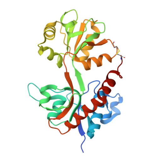
    9J8K

  • PubMed Abstract: 

    The subcellular localization of neurotransmitter receptors is strictly regulated in neurons. Changes in the trafficking of α-amino-3-hydroxy-5-methyl-4-isoxazolepropionic acid (AMPA)-type glutamate receptors (AMPARs) play an essential role in synaptic plasticity, which is the cellular basis of learning and memory. To explore receptor trafficking, genetically encoded approaches (e.g., the fusion of fluorescent proteins to receptors) are often used. However, concerns remain that genetic approaches cannot fully reproduce the receptor functions that are inherent to neurons. Herein, we report on PFQX1(AF488), a fluorescent probe for the visualization of cell-surface AMPARs without any genetic manipulation to neurons. The rapid and reversible staining features of this probe enabled snapshot imaging, which showed the accumulation of native AMPARs in dendritic spines during synaptic plasticity. Moreover, the mechanism of this synaptic accumulation, for which genetically encoded approaches have given controversial results, was revealed by integrating two chemical methods: PFQX1(AF488) and covalent chemical labeling.


  • Organizational Affiliation

    Department of Biomolecular Engineering, Graduate School of Engineering, Nagoya University, Nagoya, Aichi 464-8603, Japan.


Macromolecules
Find similar proteins by:  (by identity cutoff)  |  3D Structure
Entity ID: 1
MoleculeChains Sequence LengthOrganismDetailsImage
GluA2 ligand binding core272Rattus norvegicusMutation(s): 0 
Entity Groups  
Sequence Clusters30% Identity50% Identity70% Identity90% Identity95% Identity100% Identity
Sequence Annotations
Expand
  • Reference Sequence
Small Molecules
Ligands 3 Unique
IDChains Name / Formula / InChI Key2D Diagram3D Interactions
A1L33 (Subject of Investigation/LOI)
Query on A1L33

Download Ideal Coordinates CCD File 
B [auth A]2-(3-azanyl-6-azanylidene-4,5-disulfo-xanthen-9-yl)-5-[[1-[4-(2-hydroxy-2-oxoethyl)-2,3-bis(oxidanylidene)-7-(trifluoromethyl)-1~{H}-quinoxalin-6-yl]pyrrol-3-yl]methylcarbamoyl]benzoic acid
C37 H25 F3 N6 O14 S2
WYMZJRUHFVFSCC-YTJQMYRNSA-N
PEG
Query on PEG

Download Ideal Coordinates CCD File 
E [auth A],
F [auth A]
DI(HYDROXYETHYL)ETHER
C4 H10 O3
MTHSVFCYNBDYFN-UHFFFAOYSA-N
SO4
Query on SO4

Download Ideal Coordinates CCD File 
C [auth A],
D [auth A]
SULFATE ION
O4 S
QAOWNCQODCNURD-UHFFFAOYSA-L
Experimental Data & Validation

Experimental Data

  • Method: X-RAY DIFFRACTION
  • Resolution: 1.82 Å
  • R-Value Free:  0.200 (Depositor), 0.204 (DCC) 
  • R-Value Work:  0.169 (Depositor), 0.173 (DCC) 
  • R-Value Observed: 0.171 (Depositor) 
Space Group: P 61 2 2
Unit Cell:
Length ( Å )Angle ( ˚ )
a = 69.94α = 90
b = 69.94β = 90
c = 227.52γ = 120
Software Package:
Software NamePurpose
XDSdata reduction
XSCALEdata scaling
PHASERphasing
PHENIXrefinement

Structure Validation

View Full Validation Report


Ligand Structure Quality Assessment 


Entry History & Funding Information

Deposition Data


Funding OrganizationLocationGrant Number
Japan Society for the Promotion of Science (JSPS)Japan19H05781
Japan Society for the Promotion of Science (JSPS)Japan24H00547
Japan Society for the Promotion of Science (JSPS)Japan24H02262

Revision History  (Full details and data files)

  • Version 1.0: 2025-07-02
    Type: Initial release