9JL7 | pdb_00009jl7

Crystal structure of Actinomycin D-Doxorubicin-d(AGCCGT)2 DNA ternary complex


Experimental Data Snapshot

  • Method: X-RAY DIFFRACTION
  • Resolution: 1.52 Å
  • R-Value Free: 
    0.213 (Depositor), 0.216 (DCC) 
  • R-Value Work: 
    0.203 (Depositor), 0.204 (DCC) 
  • R-Value Observed: 
    0.204 (Depositor) 

Starting Model: experimental
View more details

wwPDB Validation   3D Report Full Report


Ligand Structure Quality Assessment 


This is version 1.1 of the entry. See complete history


Literature

Structural and Functional Insights into Targeting GCCG Sites in the EGFR Promoter by Two DNA Intercalators to Inhibit Breast Cancer Metastasis.

Chang, C.C.Li, H.J.Satange, R.Lin, S.M.Chen, T.L.Lin, C.C.Neidle, S.Hou, M.H.

(2025) J Med Chem 68: 6601-6615

  • DOI: https://doi.org/10.1021/acs.jmedchem.4c03203
  • Primary Citation of Related Structures:  
    9JL7

  • PubMed Abstract: 

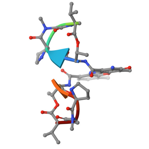
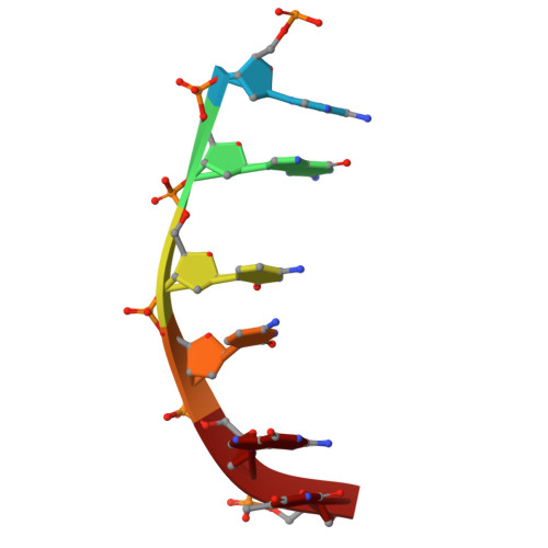
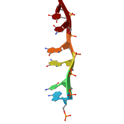
    Chemotherapeutic drugs are commonly used to treat cancers lacking targeted therapy options. However, their low specificity limits their treatment effectiveness. We report here that the cooperative binding of doxorubicin (Dox) with actinomycin D (ActD) enhances the specificity for consecutive GCCG sites in DNA. Using X-ray crystallography, we determined the crystal structure of ActD and Dox bound to d(AGCCGT) 2 DNA. ActD intercalation at the GpC site induces a novel Dox binding mode at the adjacent CpG step. This ensures a snug fit, avoids steric clashes, and enhances the specificity. Transcriptome analysis revealed that combining Dox with ActD synergistically down-regulates EGFR in TNBC cells. Additionally, it reduces EGFR promoter activity. In vivo, the combination significantly suppresses tumor growth and outperforms the standard Dox and cyclophosphamide regimen in inhibiting metastasis. This study highlights targeting the activated EGFR pathway with sequence-specific DNA-targeting drug combinations as a potential TNBC treatment.


  • Organizational Affiliation

    Graduate Institute of Biotechnology, National Chung Hsing University, Taichung 402, Taiwan.


Macromolecules

Find similar proteins by:  Sequence   |   3D Structure  

Entity ID: 3
MoleculeChains Sequence LengthOrganismDetailsImage
Actinomycin DC [auth G]11Streptomyces parvulusMutation(s): 0 
Entity Groups  
Sequence Clusters30% Identity50% Identity70% Identity90% Identity95% Identity100% Identity
Sequence Annotations
Expand
  • Reference Sequence

Find similar nucleic acids by:  Sequence   |   3D Structure  

Entity ID: 1
MoleculeChains LengthOrganismImage
DNA (5'-D(P*AP*GP*CP*CP*GP*T)-3')6synthetic construct
Sequence Annotations
Expand
  • Reference Sequence

Find similar nucleic acids by:  Sequence   |   3D Structure  

Entity ID: 2
MoleculeChains LengthOrganismImage
DNA (5'-D(P*AP*CP*GP*GP*CP*T)-3')6synthetic construct
Sequence Annotations
Expand
  • Reference Sequence
Small Molecules
Modified Residues  2 Unique
IDChains TypeFormula2D DiagramParent
MVA
Query on MVA
C [auth G]L-PEPTIDE LINKINGC6 H13 N O2VAL
SAR
Query on SAR
C [auth G]PEPTIDE LINKINGC3 H7 N O2GLY
Biologically Interesting Molecules (External Reference) 1 Unique
Experimental Data & Validation

Experimental Data

  • Method: X-RAY DIFFRACTION
  • Resolution: 1.52 Å
  • R-Value Free:  0.213 (Depositor), 0.216 (DCC) 
  • R-Value Work:  0.203 (Depositor), 0.204 (DCC) 
  • R-Value Observed: 0.204 (Depositor) 
Space Group: P 65 2 2
Unit Cell:
Length ( Å )Angle ( ˚ )
a = 27.002α = 90
b = 27.002β = 90
c = 205.993γ = 120
Software Package:
Software NamePurpose
PHENIXrefinement
HKL-2000data scaling
HKL-2000data reduction
PHENIXphasing

Structure Validation

View Full Validation Report


Ligand Structure Quality Assessment 


Entry History & Funding Information

Deposition Data


Funding OrganizationLocationGrant Number
Ministry of Science and Technology (MoST, Taiwan)Taiwan109-2628-M-005-001-MY4

Revision History  (Full details and data files)

  • Version 1.0: 2025-03-12
    Type: Initial release
  • Version 1.1: 2025-04-09
    Changes: Database references