9KK5 | pdb_00009kk5

Crystal Structure of Bovine Pancreatic Trypsin in Complex with Pyridoxal

  • Classification: HYDROLASE
  • Organism(s): Bos taurus
  • Mutation(s): No 

  • Deposited: 2024-11-12 Released: 2025-04-09 
  • Deposition Author(s): Akbar, Z., Ahmad, M.S.
  • Funding Organization(s): Not funded

Experimental Data Snapshot

  • Method: X-RAY DIFFRACTION
  • Resolution: 2.00 Å
  • R-Value Free: 
    0.232 (Depositor), 0.233 (DCC) 
  • R-Value Work: 
    0.168 (Depositor), 0.180 (DCC) 
  • R-Value Observed: 
    0.171 (Depositor) 

Starting Model: experimental
View more details

wwPDB Validation   3D Report Full Report


Ligand Structure Quality Assessment 


This is version 1.0 of the entry. See complete history


Literature

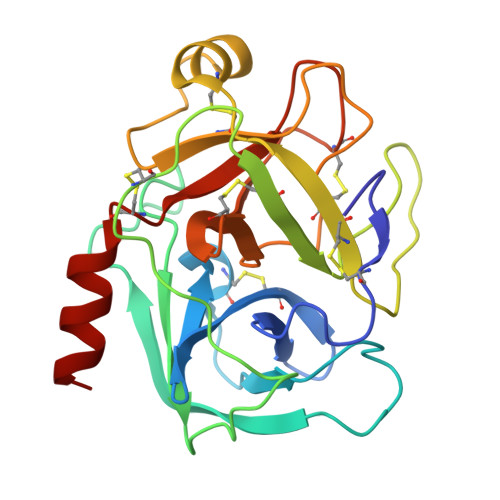
The in vitro and crystallographic studies reveal the inhibitory potential of vitamin B 6 analogues against a serine protease trypsin.

Akbar, Z.Shah, N.Mirza, S.Rasheed, S.Ahmad, M.S.

(2025) Int J Biol Macromol 308: 142433-142433

  • DOI: https://doi.org/10.1016/j.ijbiomac.2025.142433
  • Primary Citation of Related Structures:  
    9KK4, 9KK5

  • PubMed Abstract: 

    Trypsin is a representative member of serine protease family of peptidases. Its premature activation leads to develop several ailments, such as pancreatitis and colorectal cancer. The available therapies are few in numbers and possessed several side effects. Therefore, searching of new trypsin inhibitors has a great importance in drug discovery. In the current study, we have discovered the inhibitory potential of vitamin B 6 and its commercially available analogues; pyridoxine (1), pyridoxal (2), pyridoxamine (3) and pyridoxal phosphate (4), against trypsin through in vitro and crystallographic approaches. Compound 1 (pyridoxine) and 2 (pyridoxal) showed significant inhibitory potential against trypsin with the IC 50 of 234.99 ± 1.41 and 235.98 ± 1.41 μM, respectively and they did not show any significant difference in their IC 50 values (p > 0.05). The mechanistic studies reveal that both molecules showed noncompetitive mode of inhibition against trypsin enzyme. The crystallographic studies reveal the binding of compounds 1 and 2 within the S1 pocket of enzyme in two different conformations. Both conformations were stabilized with hydrogen bonding. They mostly showed interactions with Ser195, a key residue of the active site.


  • Organizational Affiliation

    H.E.J. Research Institute of Chemistry, International Center for Chemical and Biological Sciences, University of Karachi, Karachi 75270, Pakistan.


Macromolecules
Find similar proteins by:  (by identity cutoff)  |  3D Structure
Entity ID: 1
MoleculeChains Sequence LengthOrganismDetailsImage
Cationic trypsin223Bos taurusMutation(s): 0 
EC: 3.4.21.4
UniProt
Find proteins for P00760 (Bos taurus)
Explore P00760 
Go to UniProtKB:  P00760
Entity Groups  
Sequence Clusters30% Identity50% Identity70% Identity90% Identity95% Identity100% Identity
UniProt GroupP00760
Sequence Annotations
Expand
  • Reference Sequence
Experimental Data & Validation

Experimental Data

  • Method: X-RAY DIFFRACTION
  • Resolution: 2.00 Å
  • R-Value Free:  0.232 (Depositor), 0.233 (DCC) 
  • R-Value Work:  0.168 (Depositor), 0.180 (DCC) 
  • R-Value Observed: 0.171 (Depositor) 
Space Group: P 21 21 21
Unit Cell:
Length ( Å )Angle ( ˚ )
a = 54.559α = 90
b = 58.123β = 90
c = 65.626γ = 90
Software Package:
Software NamePurpose
REFMACrefinement
PROTEUM PLUSdata reduction
Aimlessdata scaling
MOLREPphasing

Structure Validation

View Full Validation Report


Ligand Structure Quality Assessment 


Entry History & Funding Information

Deposition Data


Funding OrganizationLocationGrant Number
Not fundedPakistan--

Revision History  (Full details and data files)

  • Version 1.0: 2025-04-09
    Type: Initial release