9KPY | pdb_00009kpy

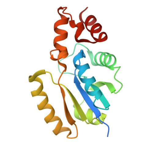
Structure of Phosphopantetheine adenylyltransferase (PPAT) from Enterobacter spp. with the expression tag bound in the substrate binding site of a neighbouring molecule at 2.20 A resolution.


Experimental Data Snapshot

  • Method: X-RAY DIFFRACTION
  • Resolution: 2.20 Å
  • R-Value Free: 
    0.267 (Depositor), 0.269 (DCC) 
  • R-Value Work: 
    0.207 (Depositor), 0.213 (DCC) 

Starting Model: experimental
View more details

wwPDB Validation   3D Report Full Report


Ligand Structure Quality Assessment 


This is version 1.1 of the entry. See complete history


Literature

Protein-tags and their fragments as potent inhibitors of enzymes: Structure of the ternary complex of phosphopantetheine adenylyltransferase from Enterobacter spp. with tag-peptides and phosphonoacetic acid at 2.20 angstrom resolution.

Ahmad, N.Kumar, V.Goel, V.K.Sharma, P.Sharma, S.Singh, T.P.

(2025) Protein Sci 34: e70216-e70216

  • DOI: https://doi.org/10.1002/pro.70216
  • Primary Citation of Related Structures:  
    9KPY

  • PubMed Abstract: 

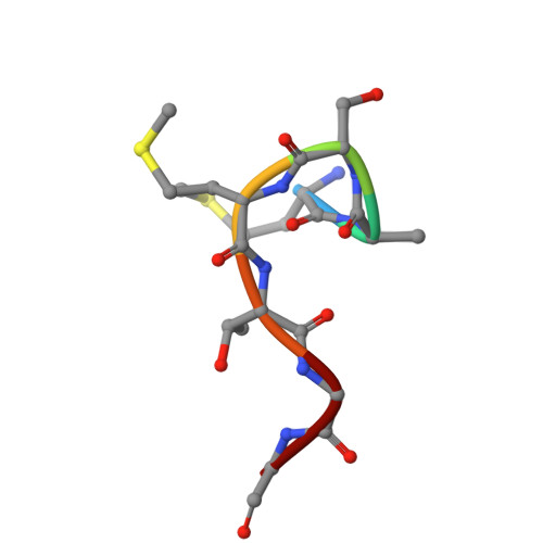
    Phosphopantetheine adenylyltransferase (PPAT) catalyzes the penultimate step of coenzyme A (CoA) biosynthesis pathway by transferring the adenylyl group from adenosine triphosphate (ATP) to 4'-phosphopantetheine (PNS), yielding 3'-dephosphocoenzyme A and pyrophosphate. In this study, the recombinant PPAT from Enterobacter spp. strain 638 (EbPPAT) was purified and co-crystallized with phosphonoacetic acid (PAE). The structure showed the presence of three homodimers AB, CD, and EF in the asymmetric unit. The 14 extra N-terminal residues (Met-14 to Ser-1, 14-mer peptide) from the expression tag were observed in Molecules B and F. These tag-peptides occupied the PNS-binding sites of adjacent Molecules A and E, respectively. Additionally, a heptapeptide (Met-14 to Gly-8) was also observed in the PNS-binding site of Molecule C. Furthermore, two PAE molecules were present in the ATP-binding sites of Molecules B, D, and F, whereas a single PAE molecule was found in Molecules A, C, and E. This showed that tag-peptides blocked the PNS-binding site while PAE blocked the ATP-binding sites. Three peptides of the tag, including 14-mer (Met-14 to Ser-1), heptapeptide (Met-14 to Gly-8) and pentapeptide (Met-14 to Thr-10) were synthesized, and their binding affinities were estimated, which showed the K D values of 5.5 × 10 -5 , 1.8 × 10 -8 , and 7.3 × 10 -9  M, respectively. PAE molecules bound to EbPPAT in the ATP-binding sites with a K D of 4.77 × 10 -4  M. This is the first structure of PPAT with peptides bound in the substrate-binding sites, indicating a novel approach to design peptide inhibitors.


  • Organizational Affiliation
    • Department of Biophysics, All India Institute of Medical Sciences, New Delhi, India.

Macromolecules
Find similar proteins by:  (by identity cutoff)  |  3D Structure
Entity ID: 1
MoleculeChains Sequence LengthOrganismDetailsImage
Phosphopantetheine adenylyltransferase
A, B, C, D, E
A, B, C, D, E, F
173Enterobacter sp. 638Mutation(s): 0 
Gene Names: coaDEnt638_0105
EC: 2.7.7.3
UniProt
Find proteins for A4W515 (Enterobacter sp. (strain 638))
Explore A4W515 
Go to UniProtKB:  A4W515
Entity Groups  
Sequence Clusters30% Identity50% Identity70% Identity90% Identity95% Identity100% Identity
UniProt GroupA4W515
Sequence Annotations
Expand
  • Reference Sequence

Find similar proteins by:  Sequence   |   3D Structure  

Entity ID: 2
MoleculeChains Sequence LengthOrganismDetailsImage
HeptapeptideG [auth H]7Enterobacter sp. 638Mutation(s): 0 
Gene Names: coaDEnt638_0105
Sequence Annotations
Expand
  • Reference Sequence
Small Molecules
Ligands 2 Unique
IDChains Name / Formula / InChI Key2D Diagram3D Interactions
PAE (Subject of Investigation/LOI)
Query on PAE

Download Ideal Coordinates CCD File 
H [auth A]
J [auth B]
K [auth B]
L [auth C]
M [auth D]
H [auth A],
J [auth B],
K [auth B],
L [auth C],
M [auth D],
N [auth D],
O [auth D],
P [auth E],
Q [auth F],
R [auth F]
PHOSPHONOACETIC ACID
C2 H5 O5 P
XUYJLQHKOGNDPB-UHFFFAOYSA-N
GOL
Query on GOL

Download Ideal Coordinates CCD File 
I [auth B]GLYCEROL
C3 H8 O3
PEDCQBHIVMGVHV-UHFFFAOYSA-N
Experimental Data & Validation

Experimental Data

  • Method: X-RAY DIFFRACTION
  • Resolution: 2.20 Å
  • R-Value Free:  0.267 (Depositor), 0.269 (DCC) 
  • R-Value Work:  0.207 (Depositor), 0.213 (DCC) 
Space Group: C 1 2 1
Unit Cell:
Length ( Å )Angle ( ˚ )
a = 137.966α = 90
b = 78.659β = 93.121
c = 107.027γ = 90
Software Package:
Software NamePurpose
REFMACrefinement
MxCuBEdata collection
XDSdata reduction
autoPROCdata scaling
MOLREPphasing
Cootmodel building

Structure Validation

View Full Validation Report


Ligand Structure Quality Assessment 


Entry History & Funding Information

Deposition Data


Funding OrganizationLocationGrant Number
Indian Council of Medical ResearchIndiaI-1251

Revision History  (Full details and data files)

  • Version 1.0: 2024-12-11
    Type: Initial release
  • Version 1.1: 2025-08-20
    Changes: Database references