9KRO | pdb_00009kro

Crystal structure of SHMT from E. faecium with mangiferin


Experimental Data Snapshot

  • Method: X-RAY DIFFRACTION
  • Resolution: 2.25 Å
  • R-Value Free: 
    0.215 (Depositor), 0.217 (DCC) 
  • R-Value Work: 
    0.175 (Depositor), 0.179 (DCC) 
  • R-Value Observed: 
    0.177 (Depositor) 

Starting Model: experimental
View more details

wwPDB Validation   3D Report Full Report


Ligand Structure Quality Assessment 


This is version 1.0 of the entry. See complete history


Literature

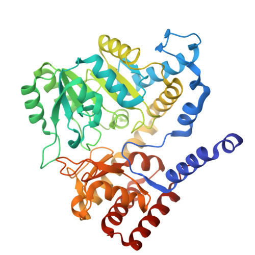
Binding of a potential antibacterial drug, mangiferin, to serine hydroxymethyltransferase from Enterococcus faecium.

Hayashi, H.Hasegawa, K.Saijo, E.Kodama, E.N.Murayama, K.

(2025) Biochem Biophys Res Commun 743: 151177-151177

  • DOI: https://doi.org/10.1016/j.bbrc.2024.151177
  • Primary Citation of Related Structures:  
    9KRO

  • PubMed Abstract: 

    Serine hydroxymethyltransferase (SHMT) plays a critical role in the 1C metabolism pathway. This pathway is involved in the synthesis of many amino and nucleic acids, and SHMT is considered a target for drugs through folate metabolism, especially for cancer and malaria. A detailed analysis of the interactions between SHMTs and drugs will greatly contribute to the development of new drugs. An anthraquinone compound was found in a compound library screening against SHMT from Enterococcus faecium (efmSHMT), from which mangiferin was implied as a compound that binds to efmSHMT. The binding assay indicated that mangiferin could bind to efmSHMT, and crystal structure analysis revealed interactions between efmSHMT and mangiferin at the binding site. Mangiferin bound to the binding site, turning the glucose moiety inward, which was supported by the docking model study. Although mangiferin does not share its molecular structure with other known inhibitors, such as pyrazolopyran-based compounds, the complex structure of the binding site did not differ much from those of other structures. The ligand binding site of efmSHMT may possess a preferred conformation.


  • Organizational Affiliation

    Division of Infectious Diseases, International Research Institute of Disaster Science, Tohoku University, 2-1, Seiryo-machi, Aoba-ku, Sendai, Miyagi, 980-8575, Japan.


Macromolecules
Find similar proteins by:  (by identity cutoff)  |  3D Structure
Entity ID: 1
MoleculeChains Sequence LengthOrganismDetailsImage
Serine hydroxymethyltransferase
A, B
417Enterococcus faeciumMutation(s): 0 
Gene Names: 
EC: 2.1.2.1
UniProt
Find proteins for A0A133CK16 (Enterococcus faecium)
Explore A0A133CK16 
Go to UniProtKB:  A0A133CK16
Entity Groups  
Sequence Clusters30% Identity50% Identity70% Identity90% Identity95% Identity100% Identity
UniProt GroupA0A133CK16
Sequence Annotations
Expand
  • Reference Sequence
Experimental Data & Validation

Experimental Data

  • Method: X-RAY DIFFRACTION
  • Resolution: 2.25 Å
  • R-Value Free:  0.215 (Depositor), 0.217 (DCC) 
  • R-Value Work:  0.175 (Depositor), 0.179 (DCC) 
  • R-Value Observed: 0.177 (Depositor) 
Space Group: P 41 21 2
Unit Cell:
Length ( Å )Angle ( ˚ )
a = 118.82α = 90
b = 118.82β = 90
c = 163.6γ = 90
Software Package:
Software NamePurpose
PHENIXrefinement
XDSdata reduction
XDSdata scaling
PHENIXphasing

Structure Validation

View Full Validation Report


Ligand Structure Quality Assessment 


Entry History & Funding Information

Deposition Data


Funding OrganizationLocationGrant Number
Japan Society for the Promotion of Science (JSPS)JapanJP23K07918

Revision History  (Full details and data files)

  • Version 1.0: 2025-03-19
    Type: Initial release