Structural basis for sensitivity and acquired resistance of fungal cap guanine-N7 methyltransferases to the antifungal antibiotic sinefungin.
Nilson, D.J., Schwer, B., Almo, S.C., Shuman, S., Ghosh, A.(2025) Nucleic Acids Res 53
- PubMed: 40682821
- DOI: https://doi.org/10.1093/nar/gkaf538
- Primary Citation of Related Structures:
9MG0, 9MG1, 9MG2, 9MG3, 9MG4, 9MG5, 9MG6 - PubMed Abstract:
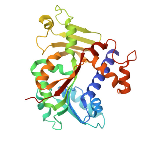
The essential enzyme messenger RNA (mRNA) (guanine-N7) methyltransferase catalyzes S-adenosylmethionine (SAM)-dependent conversion of GpppRNA ends to the m7GpppRNA cap structure characteristic of eukaryal mRNAs. The antibiotic sinefungin (SFG) is a SAM analog in which the S-CH3 sulfonium moiety of SAM is replaced by a C-NH2 amine. Available evidence indicates that the antifungal activity of SFG is exerted via inhibition of fungal cap methyltransferase Abd1. Here we report that recombinant Kluyveromyces lactis and Saccharomyces cerevisiae Abd1 are 240-fold and 485-fold more sensitive to inhibition by SFG than by the reaction product S-adenosylhomocysteine (SAH). Crystal structures of K. lactis and S. cerevisiae Abd1 as binary complexes with SAH or SFG and ternary complexes with GTP•SFG highlight how SFG makes two hydrogen bonds from its C-NH2 amine to the guanine-O6 and -N7 atoms of GTP that account for its higher affinity vis-à-vis SAH and SAM. Through a genetic screen to isolate SFG-resistant S. cerevisiae strains, a conserved tyrosine (Tyr416) that interacts with the cap guanine in Abd1 was identified as a key determinant of SFG potency. Tyr416 Abd1 variants confer SFG resistance in vitro by weakening cap-assisted SFG interactions with Abd1. Our study illuminates the basis for the exquisite SFG sensitivity of fungal cap methyltransferases.
- Department of Biochemistry, Albert Einstein College of Medicine, 1300 Morris Park Avenue, Bronx, New York, NY 10461, United States.
Organizational Affiliation: