9MS2 | pdb_00009ms2

Crystal structure of human CYP3A5 in complex with SJYHJ-111


Experimental Data Snapshot

  • Method: X-RAY DIFFRACTION
  • Resolution: 2.25 Å
  • R-Value Free: 
    0.240 (Depositor), 0.239 (DCC) 
  • R-Value Work: 
    0.198 (Depositor), 0.199 (DCC) 
  • R-Value Observed: 
    0.200 (Depositor) 

Starting Model: experimental
View more details

wwPDB Validation   3D Report Full Report


Ligand Structure Quality Assessment 


This is version 1.1 of the entry. See complete history


Literature

Decoding the selective chemical modulation of CYP3A4.

Wang, J.Nithianantham, S.Chai, S.C.Jung, Y.H.Yang, L.Ong, H.W.Li, Y.Zhang, Y.Miller, D.J.Chen, T.

(2025) Nat Commun 16: 3423-3423

  • DOI: https://doi.org/10.1038/s41467-025-58749-8
  • Primary Citation of Related Structures:  
    9BV5, 9BV6, 9BV7, 9BV8, 9BV9, 9BVA, 9BVB, 9BVC, 9MS1, 9MS2

  • PubMed Abstract: 

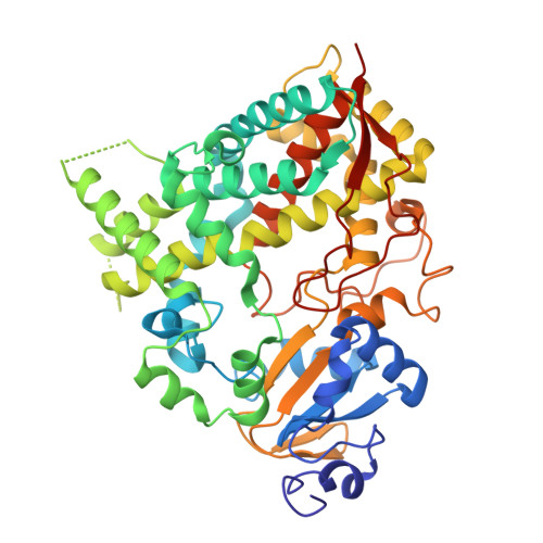
    Drug-drug interactions associate with concurrent uses of multiple medications. Cytochrome P450 (CYP) 3A4 metabolizes a large portion of marketed drugs. To maintain the efficacy of drugs metabolized by CYP3A4, pan-CYP3A inhibitors such as ritonavir are often co-administered. Although selective CYP3A4 inhibitors have greater therapeutic benefits as they avoid inhibiting unintended CYPs and undesirable clinical consequences, the high homology between CYP3A4 and CYP3A5 has hampered the development of such selective inhibitors. Here, we report a series of selective CYP3A4 inhibitors with scaffolds identified by high-throughput screening. Structural, functional, and computational analyses reveal that the differential C-terminal loop conformations and two distinct ligand binding surfaces disfavor the binding of selective CYP3A4 inhibitors to CYP3A5. Structure-guided design of compounds validates the model and yields analogs that are selective for CYP3A4 versus other major CYPs. These findings demonstrate the feasibility to selectively inhibit CYP3A4 and provide guidance for designing better CYP3A4 selective inhibitors.


  • Organizational Affiliation

    Department of Chemical Biology and Therapeutics, St. Jude Children's Research Hospital, Memphis, TN, USA.


Macromolecules
Find similar proteins by:  (by identity cutoff)  |  3D Structure
Entity ID: 1
MoleculeChains Sequence LengthOrganismDetailsImage
Cytochrome P450 3A5
A, B
480Homo sapiensMutation(s): 0 
Gene Names: CYP3A5
EC: 1.14.14.1
UniProt & NIH Common Fund Data Resources
Find proteins for P20815 (Homo sapiens)
Explore P20815 
Go to UniProtKB:  P20815
PHAROS:  P20815
GTEx:  ENSG00000106258 
Entity Groups  
Sequence Clusters30% Identity50% Identity70% Identity90% Identity95% Identity100% Identity
UniProt GroupP20815
Sequence Annotations
Expand
  • Reference Sequence
Small Molecules
Ligands 5 Unique
IDChains Name / Formula / InChI Key2D Diagram3D Interactions
HEM
Query on HEM

Download Ideal Coordinates CCD File 
C [auth A],
K [auth B]
PROTOPORPHYRIN IX CONTAINING FE
C34 H32 Fe N4 O4
KABFMIBPWCXCRK-RGGAHWMASA-L
A1BNX (Subject of Investigation/LOI)
Query on A1BNX

Download Ideal Coordinates CCD File 
D [auth A],
L [auth B]
N'-(2-chlorophenyl)-N-[(3-hydroxyphenyl)methyl]-N-[(2P)-2-(1H-imidazol-1-yl)-5-(trifluoromethyl)phenyl]urea
C24 H18 Cl F3 N4 O2
VZCCVRYFCXBWQT-UHFFFAOYSA-N
PEG
Query on PEG

Download Ideal Coordinates CCD File 
H [auth A],
Q [auth B],
R [auth B],
S [auth B]
DI(HYDROXYETHYL)ETHER
C4 H10 O3
MTHSVFCYNBDYFN-UHFFFAOYSA-N
SO4
Query on SO4

Download Ideal Coordinates CCD File 
J [auth A]SULFATE ION
O4 S
QAOWNCQODCNURD-UHFFFAOYSA-L
EDO
Query on EDO

Download Ideal Coordinates CCD File 
E [auth A]
F [auth A]
G [auth A]
I [auth A]
M [auth B]
E [auth A],
F [auth A],
G [auth A],
I [auth A],
M [auth B],
N [auth B],
O [auth B],
P [auth B]
1,2-ETHANEDIOL
C2 H6 O2
LYCAIKOWRPUZTN-UHFFFAOYSA-N
Experimental Data & Validation

Experimental Data

  • Method: X-RAY DIFFRACTION
  • Resolution: 2.25 Å
  • R-Value Free:  0.240 (Depositor), 0.239 (DCC) 
  • R-Value Work:  0.198 (Depositor), 0.199 (DCC) 
  • R-Value Observed: 0.200 (Depositor) 
Space Group: C 2 2 21
Unit Cell:
Length ( Å )Angle ( ˚ )
a = 65.085α = 90
b = 210.688β = 90
c = 153.993γ = 90
Software Package:
Software NamePurpose
PHENIXrefinement
Aimlessdata scaling
XDSdata reduction
PHENIXphasing

Structure Validation

View Full Validation Report


Ligand Structure Quality Assessment 


Entry History & Funding Information

Deposition Data


Funding OrganizationLocationGrant Number
National Institutes of Health/National Institute of General Medical Sciences (NIH/NIGMS)United StatesR35GM118041

Revision History  (Full details and data files)

  • Version 1.0: 2025-04-16
    Type: Initial release
  • Version 1.1: 2025-04-23
    Changes: Database references