9NTJ | pdb_00009ntj

Chimeric Adenosine deaminase growth factor (ADGF) in complex with pentostatin


Experimental Data Snapshot

  • Method: X-RAY DIFFRACTION
  • Resolution: 3.28 Å
  • R-Value Free: 
    0.261 (Depositor), 0.261 (DCC) 
  • R-Value Work: 
    0.210 (Depositor), 0.210 (DCC) 
  • R-Value Observed: 
    0.212 (Depositor) 

Starting Model: in silico
View more details

wwPDB Validation   3D Report Full Report


Ligand Structure Quality Assessment 


This is version 1.0 of the entry. See complete history


Literature

Structural basis for the substrate specificity of Helix pomatia AMP deaminase and a chimeric ADGF adenosine deaminase.

Kaur, G.Horton, J.R.Tzertzinis, G.Zhou, J.Schildkraut, I.Cheng, X.

(2025) J Biological Chem : 110357-110357

  • DOI: https://doi.org/10.1016/j.jbc.2025.110357
  • Primary Citation of Related Structures:  
    9NTE, 9NTF, 9NTH, 9NTI, 9NTJ, 9NTK

  • PubMed Abstract: 

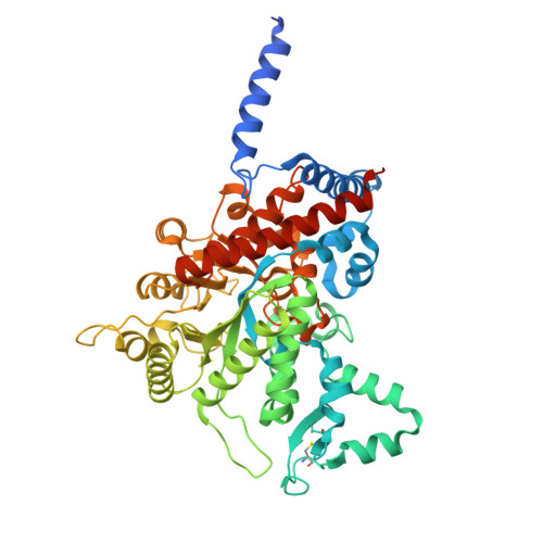
    Helix pomatia AMP deaminase (HPAMPD), an enzyme enriched in the foot muscle of the mollusk Helix pomatia, exhibits deaminase activity on adenosine-5'-monophosphate (AMP). HPAMPD is the first member of the adenosine deaminase-related growth factor (ADGF) family to prefer the nucleotideAMP over the nucleoside adenosine. To investigate the substrate selectivity of HPAMPD, we determined its structure in both the apo form and in complex with the adenosine analogs pentostatin and pentostatin-5'-monophosphate. Structurally, HPAMPD adopts a fold similar to human ADA2, an ADGF family member. HPAMPD has acquired the ability to interact with the 5'-monophosphate group of AMP through polar and charged residues located in three key structural elements: (1) the loop immediately following strand β1; (2) the loop between helices αH and αI; and (3) the end of strand β5 and its adjacent loop. We engineered a chimeric deaminase by integrating these elements from HPAMPD into another related mollusk nucleoside adenosine deaminase, Aplysia ADGF. The chimeric enzyme efficiently deaminates AMP, demonstrating a gained substrate specificity, while retaining the adenosine deamination activity of Aplysia ADGF. The phosphate-binding feature of HPAMPD is a hallmark of nucleotide deaminases, conserved among AMP and N6-methyl-AMP (6mAMP) deaminases. We discuss the human adenosine deaminases each with distinct substrate specificities for the nucleoside, the nucleotide (AMP), and its methylated form, 6mAMP.


  • Organizational Affiliation

    Department of Epigenetics and Molecular Carcinogenesis, The University of Texas MD Anderson Cancer Center, Houston, TX 77030, USA.


Macromolecules
Find similar proteins by:  (by identity cutoff)  |  3D Structure
Entity ID: 1
MoleculeChains Sequence LengthOrganismDetailsImage
Adenosine deaminase AGSA,Adenosine deaminase AGSA,AMP deaminase
A, B
536Aplysia californicaHelix pomatiaMutation(s): 0 
EC: 3.5.4.4
UniProt
Find proteins for P15287 (Aplysia californica)
Explore P15287 
Go to UniProtKB:  P15287
Entity Groups  
Sequence Clusters30% Identity50% Identity70% Identity90% Identity95% Identity100% Identity
UniProt GroupP15287
Glycosylation
Glycosylation Sites: 4
Sequence Annotations
Expand
  • Reference Sequence
Oligosaccharides

Help

Entity ID: 2
MoleculeChains Length2D Diagram Glycosylation3D Interactions
alpha-D-mannopyranose-(1-6)-beta-D-mannopyranose-(1-4)-2-acetamido-2-deoxy-beta-D-glucopyranose-(1-4)-2-acetamido-2-deoxy-beta-D-glucopyranose
C
4N-Glycosylation
Glycosylation Resources
GlyTouCan:  G22573RC
GlyCosmos:  G22573RC
GlyGen:  G22573RC
Small Molecules
Ligands 4 Unique
IDChains Name / Formula / InChI Key2D Diagram3D Interactions
DCF (Subject of Investigation/LOI)
Query on DCF

Download Ideal Coordinates CCD File 
K [auth A],
T [auth B]
2'-DEOXYCOFORMYCIN
C11 H16 N4 O4
FPVKHBSQESCIEP-JQCXWYLXSA-N
NAG
Query on NAG

Download Ideal Coordinates CCD File 
D [auth A]
E [auth A]
F [auth A]
L [auth B]
M [auth B]
D [auth A],
E [auth A],
F [auth A],
L [auth B],
M [auth B],
N [auth B],
O [auth B]
2-acetamido-2-deoxy-beta-D-glucopyranose
C8 H15 N O6
OVRNDRQMDRJTHS-FMDGEEDCSA-N
ZN
Query on ZN

Download Ideal Coordinates CCD File 
J [auth A],
S [auth B]
ZINC ION
Zn
PTFCDOFLOPIGGS-UHFFFAOYSA-N
EDO
Query on EDO

Download Ideal Coordinates CCD File 
G [auth A]
H [auth A]
I [auth A]
P [auth B]
Q [auth B]
G [auth A],
H [auth A],
I [auth A],
P [auth B],
Q [auth B],
R [auth B]
1,2-ETHANEDIOL
C2 H6 O2
LYCAIKOWRPUZTN-UHFFFAOYSA-N
Experimental Data & Validation

Experimental Data

  • Method: X-RAY DIFFRACTION
  • Resolution: 3.28 Å
  • R-Value Free:  0.261 (Depositor), 0.261 (DCC) 
  • R-Value Work:  0.210 (Depositor), 0.210 (DCC) 
  • R-Value Observed: 0.212 (Depositor) 
Space Group: P 21 21 21
Unit Cell:
Length ( Å )Angle ( ˚ )
a = 67.349α = 90
b = 109.609β = 90
c = 171.555γ = 90
Software Package:
Software NamePurpose
PHENIXrefinement
HKL-2000data reduction
SCALEPACKdata scaling
PHENIXphasing

Structure Validation

View Full Validation Report


Ligand Structure Quality Assessment 


Entry History & Funding Information

Deposition Data


Funding OrganizationLocationGrant Number
National Institutes of Health/National Institute of General Medical Sciences (NIH/NIGMS)United StatesR35GM134744
Cancer Prevention and Research Institute of Texas (CPRIT)United StatesRR160029

Revision History  (Full details and data files)

  • Version 1.0: 2025-06-25
    Type: Initial release