9R8C | pdb_00009r8c

CRYSTAL STRUCTURE OF WILD-TYPE PLK1 KINASE DOMAIN IN COMPLEX WITH THIAZOLIDINONE INHIBITOR COMPOUND 1 AND A SELECTIVE DARPIN


Experimental Data Snapshot

  • Method: X-RAY DIFFRACTION
  • Resolution: 2.24 Å
  • R-Value Free: 
    0.236 (Depositor), 0.235 (DCC) 
  • R-Value Work: 
    0.188 (Depositor), 0.193 (DCC) 

Starting Model: experimental
View more details

wwPDB Validation   3D Report Full Report


Ligand Structure Quality Assessment 


This is version 1.1 of the entry. See complete history


Literature

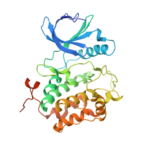
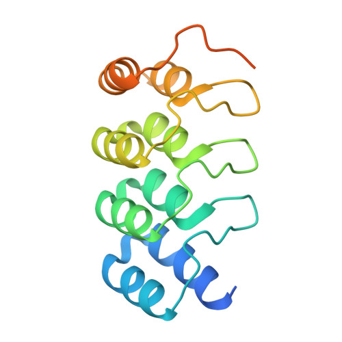
Polo-like kinase 1-inhibitor co-complex structures via the surface-entropy reduction approach and a DARPin-assisted approach.

Eberspaecher, U.Schmitz, A.A.Siemeister, G.Bomer, U.Bandeiras, T.M.Matias, P.M.Schulze, V.K.Hillig, R.C.

(2025) Acta Crystallogr D Struct Biol 81: 718-733

  • DOI: https://doi.org/10.1107/S2059798325009325
  • Primary Citation of Related Structures:  
    9R1W, 9R1X, 9R1Y, 9R8B, 9R8C

  • PubMed Abstract: 

    Polo-like kinase 1 (PLK1) is a major regulator of cell division and has been pursued as a drug target for cancer therapy for a long time. Crystallization of the kinase domain has proven to be exceptionally challenging. Previously, we published a crystallization approach using a PLK1-specific designed ankyrin-repeat protein (DARPin) as a crystallization facilitator. Here, we report an alternative route: crystallization was successful after the introduction of a double mutation which reduced surface entropy and enabled the formation of a new crystal contact. This new PLK1 crystallization system was used to determine the first co-complex crystal structure of the Bayer thiazolidinone lead series, as well as crystal structures with representatives of two competitor inhibitor series. The molecular binding modes of these three inhibitors are analysed and discussed, and the surface-entropy reduction approach is compared with the surface modifications employed by us and others to enable the crystallization of PLK1.


  • Organizational Affiliation
    • Research and Development, Pharmaceuticals, Bayer AG, Muellerstrasse 178, 13353 Berlin, Germany.

Macromolecules
Find similar proteins by:  (by identity cutoff)  |  3D Structure
Entity ID: 1
MoleculeChains Sequence LengthOrganismDetailsImage
Serine/threonine-protein kinase PLK1
A, B
315Homo sapiensMutation(s): 0 
Gene Names: PLK1PLK
EC: 2.7.11.21
UniProt & NIH Common Fund Data Resources
Find proteins for P53350 (Homo sapiens)
Explore P53350 
Go to UniProtKB:  P53350
PHAROS:  P53350
GTEx:  ENSG00000166851 
Entity Groups  
Sequence Clusters30% Identity50% Identity70% Identity90% Identity95% Identity100% Identity
UniProt GroupP53350
Sequence Annotations
Expand
  • Reference Sequence
Find similar proteins by:  (by identity cutoff)  |  3D Structure
Entity ID: 2
MoleculeChains Sequence LengthOrganismDetailsImage
DESIGNED ANKYRIN REPEAT PROTEIN 3H10
C, D
167Homo sapiensMutation(s): 0 
Entity Groups  
Sequence Clusters30% Identity50% Identity70% Identity90% Identity95% Identity100% Identity
Sequence Annotations
Expand
  • Reference Sequence
Experimental Data & Validation

Experimental Data

  • Method: X-RAY DIFFRACTION
  • Resolution: 2.24 Å
  • R-Value Free:  0.236 (Depositor), 0.235 (DCC) 
  • R-Value Work:  0.188 (Depositor), 0.193 (DCC) 
Space Group: P 21 21 21
Unit Cell:
Length ( Å )Angle ( ˚ )
a = 62.605α = 90
b = 135.302β = 90
c = 136.7γ = 90
Software Package:
Software NamePurpose
REFMACrefinement
XDSdata reduction
SCALAdata scaling
PHASERphasing

Structure Validation

View Full Validation Report


Ligand Structure Quality Assessment 


Entry History & Funding Information

Deposition Data


Funding OrganizationLocationGrant Number
Not funded--

Revision History  (Full details and data files)

  • Version 1.0: 2025-11-26
    Type: Initial release
  • Version 1.1: 2025-12-10
    Changes: Database references