1YDP | pdb_00001ydp

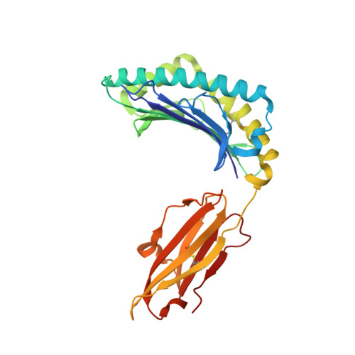
1.9A crystal structure of HLA-G


Experimental Data Snapshot

  • Method: X-RAY DIFFRACTION
  • Resolution: 1.90 Å
  • R-Value Free: 
    0.264 (Depositor) 
  • R-Value Work: 
    0.235 (Depositor) 

wwPDB Validation   3D Report Full Report


This is version 1.5 of the entry. See complete history


Literature

Crystal structure of HLA-G: A nonclassical MHC class I molecule expressed at the fetal-maternal interface

Clements, C.S.Kjer-Nielsen, L.Kostenko, L.Hoare, H.L.Dunstone, M.A.Moses, E.Freed, K.Brooks, A.G.Rossjohn, J.McCluskey, J.

(2005) Proc Natl Acad Sci U S A 102: 3360-3365

  • DOI: https://doi.org/10.1073/pnas.0409676102
  • Primary Citation of Related Structures:  
    1YDP

  • PubMed Abstract: 

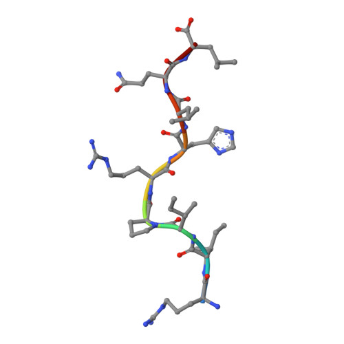
    HLA-G is a nonclassical major histocompatibility complex class I (MHC-I) molecule that is primarily expressed at the fetal-maternal interface, where it is thought to play a role in protecting the fetus from the maternal immune response. HLA-G binds a limited repertoire of peptides and interacts with the inhibitory leukocyte Ig-like receptors LIR-1 and LIR-2 and possibly with certain natural killer cell receptors. To gain further insights into HLA-G function, we determined the 1.9-A structure of a monomeric HLA-G complexed to a natural endogenous peptide ligand from histone H2A (RIIPRHLQL). An extensive network of contacts between the peptide and the antigen-binding cleft reveal a constrained mode of binding reminiscent of the nonclassical HLA-E molecule, thereby providing a structural basis for the limited peptide repertoire of HLA-G. The alpha3 domain of HLA-G, a candidate binding site for the LIR-1 and -2 inhibitory receptors, is structurally distinct from the alpha3 domains of classical MHC-I molecules, providing a rationale for the observed affinity differences for these ligands. The structural data suggest a head-to-tail mode of dimerization, mediated by an intermolecular disulfide bond, that is consistent with the observation of HLA-G dimers on the cell surface.


  • Organizational Affiliation
    • Protein Crystallography Unit, Monash Centre for Synchrotron Science, Department of Biochemistry and Molecular Biology, School of Biomedical Sciences, Monash University, Clayton, Victoria 3800, Australia.

Macromolecules
Find similar proteins by:  (by identity cutoff)  |  3D Structure
Entity ID: 1
MoleculeChains Sequence LengthOrganismDetailsImage
MHC class I antigen275Homo sapiensMutation(s): 1 
UniProt & NIH Common Fund Data Resources
Find proteins for P17693 (Homo sapiens)
Explore P17693 
Go to UniProtKB:  P17693
PHAROS:  P17693
GTEx:  ENSG00000204632 
Entity Groups  
Sequence Clusters30% Identity50% Identity70% Identity90% Identity95% Identity100% Identity
UniProt GroupP17693
Sequence Annotations
Expand
  • Reference Sequence
Find similar proteins by:  (by identity cutoff)  |  3D Structure
Entity ID: 2
MoleculeChains Sequence LengthOrganismDetailsImage
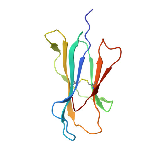
Beta-2-microglobulin100Homo sapiensMutation(s): 0 
UniProt & NIH Common Fund Data Resources
Find proteins for P61769 (Homo sapiens)
Explore P61769 
Go to UniProtKB:  P61769
PHAROS:  P61769
GTEx:  ENSG00000166710 
Entity Groups  
Sequence Clusters30% Identity50% Identity70% Identity90% Identity95% Identity100% Identity
UniProt GroupP61769
Sequence Annotations
Expand
  • Reference Sequence

Find similar proteins by:  Sequence   |   3D Structure  

Entity ID: 3
MoleculeChains Sequence LengthOrganismDetailsImage
histone 2a peptideC [auth P]9synthetic constructMutation(s): 0 
UniProt & NIH Common Fund Data Resources
Find proteins for P16104 (Homo sapiens)
Explore P16104 
Go to UniProtKB:  P16104
PHAROS:  P16104
GTEx:  ENSG00000188486 
Entity Groups  
UniProt GroupP16104
Sequence Annotations
Expand
  • Reference Sequence
Experimental Data & Validation

Experimental Data

  • Method: X-RAY DIFFRACTION
  • Resolution: 1.90 Å
  • R-Value Free:  0.264 (Depositor) 
  • R-Value Work:  0.235 (Depositor) 
Space Group: P 32 2 1
Unit Cell:
Length ( Å )Angle ( ˚ )
a = 77.15α = 90
b = 77.15β = 90
c = 151.72γ = 120
Software Package:
Software NamePurpose
CNSrefinement

Structure Validation

View Full Validation Report


Entry History 

Deposition Data

Revision History  (Full details and data files)

  • Version 1.0: 2005-03-08
    Type: Initial release
  • Version 1.1: 2008-04-30
    Changes: Version format compliance
  • Version 1.2: 2011-07-13
    Changes: Version format compliance
  • Version 1.3: 2018-07-25
    Changes: Data collection, Source and taxonomy, Structure summary
  • Version 1.4: 2021-11-10
    Changes: Database references, Derived calculations
  • Version 1.5: 2024-10-23
    Changes: Data collection, Structure summary