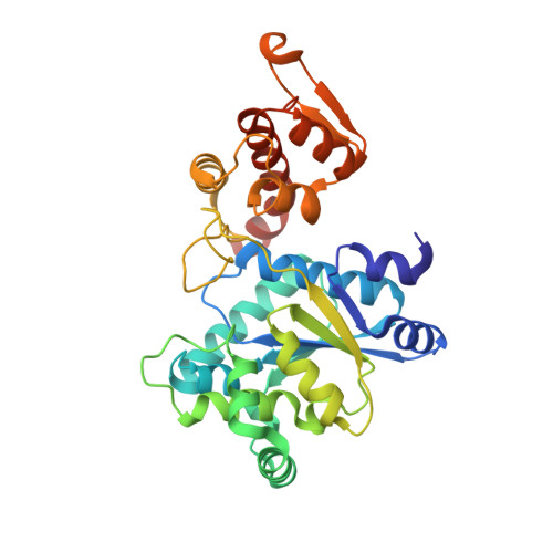
2AG6 | pdb_00002ag6

Crystal structure of p-bromo-l-phenylalanine-tRNA sythetase in complex with p-bromo-l-phenylalanine


Experimental Data Snapshot

  • Method: X-RAY DIFFRACTION
  • Resolution: 1.90 Å
  • R-Value Free: 
    0.247 (Depositor), 0.220 (DCC) 
  • R-Value Work: 
    0.209 (Depositor), 0.220 (DCC) 
  • R-Value Observed: 
    0.211 (Depositor) 

Starting Model: experimental
View more details

wwPDB Validation   3D Report Full Report


Ligand Structure Quality Assessment 


This is version 1.6 of the entry. See complete history


Literature

Structural plasticity of an aminoacyl-tRNA synthetase active site

Turner, J.M.Graziano, J.Spraggon, G.Schultz, P.G.

(2006) Proc Natl Acad Sci U S A 103: 6483-6488

  • DOI: https://doi.org/10.1073/pnas.0601756103
  • Primary Citation of Related Structures:  
    1ZH0, 2AG6

  • PubMed Abstract: 

    Recently, tRNA aminoacyl-tRNA synthetase pairs have been evolved that allow one to genetically encode a large array of unnatural amino acids in both prokaryotic and eukaryotic organisms. We have determined the crystal structures of two substrate-bound Methanococcus jannaschii tyrosyl aminoacyl-tRNA synthetases that charge the unnatural amino acids p-bromophenylalanine and 3-(2-naphthyl)alanine (NpAla). A comparison of these structures with the substrate-bound WT synthetase, as well as a mutant synthetase that charges p-acetylphenylalanine, shows that altered specificity is due to both side-chain and backbone rearrangements within the active site that modify hydrogen bonds and packing interactions with substrate, as well as disrupt the alpha8-helix, which spans the WT active site. The high degree of structural plasticity that is observed in these aminoacyl-tRNA synthetases is rarely found in other mutant enzymes with altered specificities and provides an explanation for the surprising adaptability of the genetic code to novel amino acids.


  • Organizational Affiliation
    • Department of Chemistry and The Skaggs Institute for Chemical Biology, The Scripps Research Institute, 10550 North Torrey Pines Road, La Jolla, CA 92037, USA.

Macromolecules
Find similar proteins by:  (by identity cutoff)  |  3D Structure
Entity ID: 1
MoleculeChains Sequence LengthOrganismDetailsImage
Tyrosyl-tRNA synthetase314Methanocaldococcus jannaschiiMutation(s): 5 
Gene Names: tyrS
EC: 6.1.1.1
UniProt
Find proteins for Q57834 (Methanocaldococcus jannaschii (strain ATCC 43067 / DSM 2661 / JAL-1 / JCM 10045 / NBRC 100440))
Explore Q57834 
Go to UniProtKB:  Q57834
Entity Groups  
Sequence Clusters30% Identity50% Identity70% Identity90% Identity95% Identity100% Identity
UniProt GroupQ57834
Sequence Annotations
Expand
  • Reference Sequence
Small Molecules
Ligands 1 Unique
IDChains Name / Formula / InChI Key2D Diagram3D Interactions
4BF
Query on 4BF

Download Ideal Coordinates CCD File 
B [auth A]4-BROMO-L-PHENYLALANINE
C9 H10 Br N O2
PEMUHKUIQHFMTH-QMMMGPOBSA-N
Experimental Data & Validation

Experimental Data

  • Method: X-RAY DIFFRACTION
  • Resolution: 1.90 Å
  • R-Value Free:  0.247 (Depositor), 0.220 (DCC) 
  • R-Value Work:  0.209 (Depositor), 0.220 (DCC) 
  • R-Value Observed: 0.211 (Depositor) 
Space Group: P 43 21 2
Unit Cell:
Length ( Å )Angle ( ˚ )
a = 102.719α = 90
b = 102.719β = 90
c = 70.598γ = 90
Software Package:
Software NamePurpose
REFMACrefinement
HKL-2000data reduction
SCALEPACKdata scaling

Structure Validation

View Full Validation Report


Ligand Structure Quality Assessment 


Entry History 

Deposition Data

Revision History  (Full details and data files)

  • Version 1.0: 2006-04-04
    Type: Initial release
  • Version 1.1: 2008-04-02
    Changes: Version format compliance
  • Version 1.2: 2011-07-13
    Changes: Advisory, Derived calculations, Version format compliance
  • Version 1.3: 2017-10-11
    Changes: Refinement description
  • Version 1.4: 2021-10-20
    Changes: Database references, Derived calculations
  • Version 1.5: 2023-08-23
    Changes: Data collection, Refinement description
  • Version 1.6: 2023-11-15
    Changes: Data collection