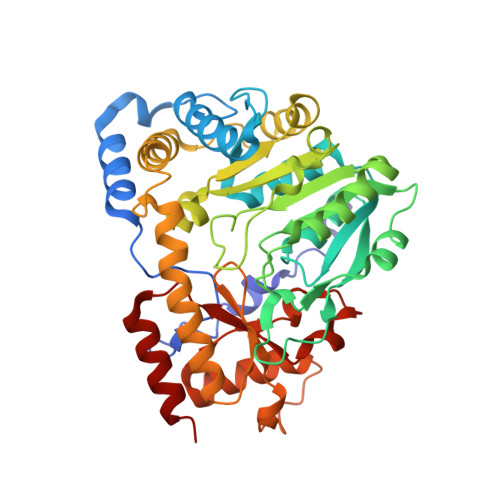
3E2Y | pdb_00003e2y

Crystal structure of mouse kynurenine aminotransferase III in complex with glutamine


Experimental Data Snapshot

  • Method: X-RAY DIFFRACTION
  • Resolution: 2.26 Å
  • R-Value Free: 
    0.221 (Depositor), 0.220 (DCC) 
  • R-Value Work: 
    0.175 (Depositor), 0.170 (DCC) 
  • R-Value Observed: 
    0.177 (Depositor) 

Starting Model: experimental
View more details

wwPDB Validation   3D Report Full Report


Ligand Structure Quality Assessment 


This is version 1.3 of the entry. See complete history

Re-refinement Note

A newer entry is available that reflects an alternative modeling of the original data: 5VEQ


Literature

Correction for Han et al., "Biochemical and Structural Properties of Mouse Kynurenine Aminotransferase III".

Han, Q.Robinson, H.Cai, T.Tagle, D.A.Li, J.

(2018) Mol Cell Biol 38

  • DOI: https://doi.org/10.1128/MCB.00099-18
  • Primary Citation of Related Structures:  
    3E2F, 3E2Y, 3E2Z

  • Organizational Affiliation
    • Department of Biochemistry, Virginia Tech, Blacksburg, Virginia 24061.

Macromolecules
Find similar proteins by:  (by identity cutoff)  |  3D Structure
Entity ID: 1
MoleculeChains Sequence LengthOrganismDetailsImage
Kynurenine-oxoglutarate transaminase 3
A, B
410Mus musculusMutation(s): 0 
Gene Names: Ccbl2Kat3
EC: 2.6.1.7 (PDB Primary Data), 4.4.1.13 (PDB Primary Data), 2.6.1.63 (UniProt)
UniProt
Find proteins for Q71RI9 (Mus musculus)
Explore Q71RI9 
Go to UniProtKB:  Q71RI9
Entity Groups  
Sequence Clusters30% Identity50% Identity70% Identity90% Identity95% Identity100% Identity
UniProt GroupQ71RI9
Sequence Annotations
Expand
  • Reference Sequence
Small Molecules
Ligands 3 Unique
IDChains Name / Formula / InChI Key2D Diagram3D Interactions
PMP
Query on PMP

Download Ideal Coordinates CCD File 
D [auth A],
G [auth B]
4'-DEOXY-4'-AMINOPYRIDOXAL-5'-PHOSPHATE
C8 H13 N2 O5 P
ZMJGSOSNSPKHNH-UHFFFAOYSA-N
GLN
Query on GLN

Download Ideal Coordinates CCD File 
C [auth A],
H [auth B]
GLUTAMINE
C5 H10 N2 O3
ZDXPYRJPNDTMRX-VKHMYHEASA-N
GOL
Query on GOL

Download Ideal Coordinates CCD File 
E [auth A],
F [auth A],
I [auth B],
J [auth B]
GLYCEROL
C3 H8 O3
PEDCQBHIVMGVHV-UHFFFAOYSA-N
Experimental Data & Validation

Experimental Data

  • Method: X-RAY DIFFRACTION
  • Resolution: 2.26 Å
  • R-Value Free:  0.221 (Depositor), 0.220 (DCC) 
  • R-Value Work:  0.175 (Depositor), 0.170 (DCC) 
  • R-Value Observed: 0.177 (Depositor) 
Space Group: P 43 21 2
Unit Cell:
Length ( Å )Angle ( ˚ )
a = 91.567α = 90
b = 91.567β = 90
c = 232.536γ = 90
Software Package:
Software NamePurpose
REFMACrefinement
HKL-2000data collection
DENZOdata reduction
SCALAdata scaling
MOLREPphasing

Structure Validation

View Full Validation Report


Ligand Structure Quality Assessment 


Entry History 

Deposition Data

Revision History  (Full details and data files)

  • Version 1.0: 2008-12-30
    Type: Initial release
  • Version 1.1: 2011-07-13
    Changes: Non-polymer description, Version format compliance
  • Version 1.2: 2018-08-01
    Changes: Advisory, Data collection, Database references
  • Version 1.3: 2023-08-30
    Changes: Data collection, Database references, Derived calculations, Refinement description