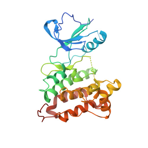
3EL7 | pdb_00003el7

Crystal structure of c-Src in complex with pyrazolopyrimidine 3


Experimental Data Snapshot

  • Method: X-RAY DIFFRACTION
  • Resolution: 2.80 Å
  • R-Value Free: 
    0.288 (Depositor), 0.270 (DCC) 
  • R-Value Work: 
    0.220 (Depositor), 0.220 (DCC) 
  • R-Value Observed: 
    0.220 (Depositor) 

Starting Model: experimental
View more details

wwPDB Validation   3D Report Full Report


Ligand Structure Quality Assessment 


This is version 1.3 of the entry. See complete history


Literature

Small molecule recognition of c-Src via the Imatinib-binding conformation.

Dar, A.C.Lopez, M.S.Shokat, K.M.

(2008) Chem Biol 15: 1015-1022

  • DOI: https://doi.org/10.1016/j.chembiol.2008.09.007
  • Primary Citation of Related Structures:  
    3EL7, 3EL8

  • PubMed Abstract: 

    The cancer drug, Imatinib, is a selective Abl kinase inhibitor that does not inhibit the closely related kinase c-Src. This one drug and its ability to selectively inhibit Abl over c-Src has been a guiding principle in virtually all kinase drug discovery efforts in the last 15 years. A prominent hypothesis explaining the selectivity of Imatinib is that Abl has an intrinsic ability to adopt an inactive conformation (termed DFG-out), whereas c-Src appears to pay a high intrinsic energetic penalty for adopting this conformation, effectively excluding Imatinib from its ATP pocket. This explanation of the difference in binding affinity of Imatinib for Abl versus c-Src makes the striking prediction that it would not be possible to design an inhibitor that binds to the DFG-out conformation of c-Src with high affinity. We report the discovery of a series of such inhibitors. We use structure-activity relationships and X-ray crystallography to confirm our findings. These studies suggest that small molecules are capable of inducing the generally unfavorable DFG-out conformation in c-Src. Structural comparison between c-Src in complex with these inhibitors allows us to speculate on the differential selectivity of Imatinib for c-Src and Abl.


  • Organizational Affiliation
    • Department of Cellular and Molecular Pharmacology, Howard Hughes Medical Institute, Genentech Hall, 600 16(th) Street, University of California, San Francisco, San Francisco, CA 94158, USA.

Macromolecules
Find similar proteins by:  (by identity cutoff)  |  3D Structure
Entity ID: 1
MoleculeChains Sequence LengthOrganismDetailsImage
Proto-oncogene tyrosine-protein kinase Src286Gallus gallusMutation(s): 0 
Gene Names: SRC
EC: 2.7.10.2
UniProt
Find proteins for P00523 (Gallus gallus)
Explore P00523 
Go to UniProtKB:  P00523
Entity Groups  
Sequence Clusters30% Identity50% Identity70% Identity90% Identity95% Identity100% Identity
UniProt GroupP00523
Sequence Annotations
Expand
  • Reference Sequence
Small Molecules
Ligands 1 Unique
IDChains Name / Formula / InChI Key2D Diagram3D Interactions
PD3
Query on PD3

Download Ideal Coordinates CCD File 
B [auth A]1-{3-[(4-amino-1-cyclopentyl-1H-pyrazolo[3,4-d]pyrimidin-3-yl)methyl]phenyl}-3-[3-(trifluoromethyl)phenyl]urea
C25 H24 F3 N7 O
RKZUINFXRREENO-UHFFFAOYSA-N
Experimental Data & Validation

Experimental Data

  • Method: X-RAY DIFFRACTION
  • Resolution: 2.80 Å
  • R-Value Free:  0.288 (Depositor), 0.270 (DCC) 
  • R-Value Work:  0.220 (Depositor), 0.220 (DCC) 
  • R-Value Observed: 0.220 (Depositor) 
Space Group: P 1 21 1
Unit Cell:
Length ( Å )Angle ( ˚ )
a = 42.36α = 90
b = 63.13β = 91.9
c = 56.05γ = 90
Software Package:
Software NamePurpose
XDSdata scaling
PHASERphasing
CNSrefinement
XDSdata reduction

Structure Validation

View Full Validation Report


Ligand Structure Quality Assessment 


Entry History 

Deposition Data

Revision History  (Full details and data files)

  • Version 1.0: 2008-10-28
    Type: Initial release
  • Version 1.1: 2011-07-13
    Changes: Version format compliance
  • Version 1.2: 2011-12-07
    Changes: Database references
  • Version 1.3: 2023-08-30
    Changes: Data collection, Database references, Derived calculations, Refinement description