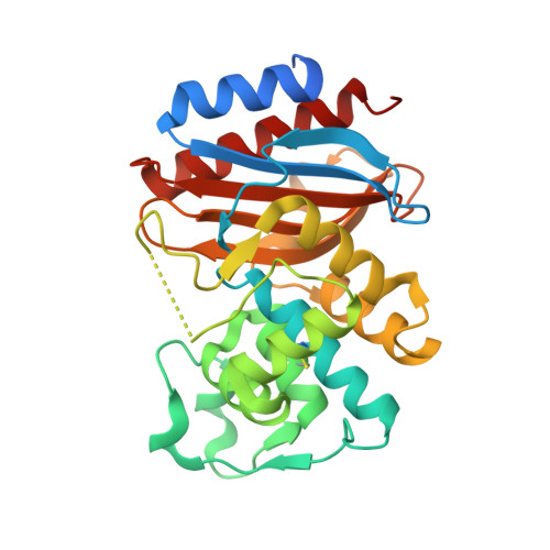
Ligand-dependent disorder of the Omega loop observed in extended-spectrum SHV-type beta-lactamase.
Sampson, J.M., Ke, W., Bethel, C.R., Pagadala, S.R., Nottingham, M.D., Bonomo, R.A., Buynak, J.D., van den Akker, F.(2011) Antimicrob Agents Chemother 55: 2303-2309
- PubMed: 21357298
- DOI: https://doi.org/10.1128/AAC.01360-10
- Primary Citation of Related Structures:
3OPH, 3OPL, 3OPP, 3OPR - PubMed Abstract:
Among Gram-negative bacteria, resistance to β-lactams is mediated primarily by β-lactamases (EC 3.2.6.5), periplasmic enzymes that inactivate β-lactam antibiotics. Substitutions at critical amino acid positions in the class A β-lactamase families result in enzymes that can hydrolyze extended-spectrum cephalosporins, thus demonstrating an "extended-spectrum" β-lactamase (ESBL) phenotype. Using SHV ESBLs with substitutions in the Ω loop (R164H and R164S) as target enzymes to understand this enhanced biochemical capability and to serve as a basis for novel β-lactamase inhibitor development, we determined the spectra of activity and crystal structures of these variants. We also studied the inactivation of the R164H and R164S mutants with tazobactam and SA2-13, a unique β-lactamase inhibitor that undergoes a distinctive reaction chemistry in the active site. We noted that the reduced Ki values for the R164H and R164S mutants with SA2-13 are comparable to those with tazobactam (submicromolar). The apo enzyme crystal structures of the R164H and R164S SHV variants revealed an ordered Ω loop architecture that became disordered when SA2-13 was bound. Important structural alterations that result from the binding of SA2-13 explain the enhanced susceptibility of these ESBL enzymes to this inhibitor and highlight ligand-dependent Ω loop flexibility as a mechanism for accommodating and hydrolyzing β-lactam substrates.
- Department of Biochemistry, Case Western University School of Medicine, Research Service, Louis Stokes Cleveland Department of Veterans Affairs Medical Center, Cleveland, OH 44106, USA.
Organizational Affiliation: