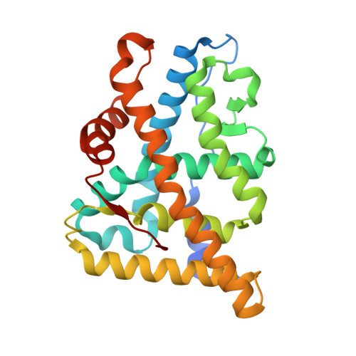
Structural basis for agonism and antagonism for a set of chemically related progesterone receptor modulators.
Lusher, S.J., Raaijmakers, H.C., Vu-Pham, D., Dechering, K., Lam, T.W., Brown, A.R., Hamilton, N.M., Nimz, O., Bosch, R., McGuire, R., Oubrie, A., de Vlieg, J.(2011) J Biological Chem 286: 35079-35086
- PubMed: 21849509
- DOI: https://doi.org/10.1074/jbc.M111.273029
- Primary Citation of Related Structures:
3ZR7, 3ZRA, 3ZRB - PubMed Abstract:
The progesterone receptor is able to bind to a large number and variety of ligands that elicit a broad range of transcriptional responses ranging from full agonism to full antagonism and numerous mixed profiles inbetween. We describe here two new progesterone receptor ligand binding domain x-ray structures bound to compounds from a structurally related but functionally divergent series, which show different binding modes corresponding to their agonistic or antagonistic nature. In addition, we present a third progesterone receptor ligand binding domain dimer bound to an agonist in monomer A and an antagonist in monomer B, which display binding modes in agreement with the earlier observation that agonists and antagonists from this series adopt different binding modes.
- Department of Molecular Design and Informatics, DMPK, MSD, PO Box 20, 5340 BH Oss, The Netherlands. scott.lusher@merck.com
Organizational Affiliation: