4JE4 | pdb_00004je4

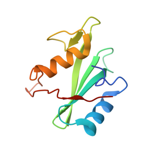
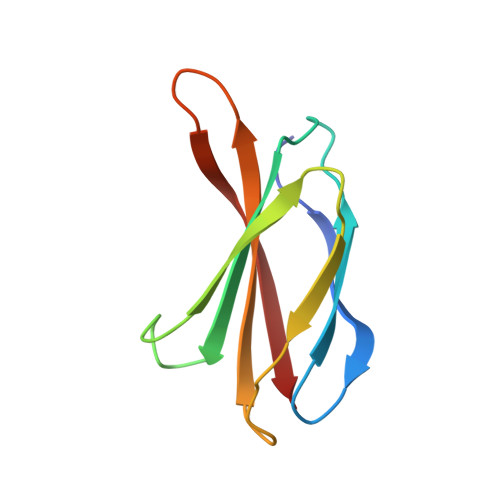
Crystal Structure of Monobody NSa1/SHP2 N-SH2 Domain Complex


Experimental Data Snapshot

  • Method: X-RAY DIFFRACTION
  • Resolution: 2.31 Å
  • R-Value Free: 
    0.255 (Depositor), 0.250 (DCC) 
  • R-Value Work: 
    0.204 (Depositor), 0.200 (DCC) 
  • R-Value Observed: 
    0.206 (Depositor) 

wwPDB Validation   3D Report Full Report


This is version 1.4 of the entry. See complete history


Literature

Dissection of the BCR-ABL signaling network using highly specific monobody inhibitors to the SHP2 SH2 domains.

Sha, F.Gencer, E.B.Georgeon, S.Koide, A.Yasui, N.Koide, S.Hantschel, O.

(2013) Proc Natl Acad Sci U S A 110: 14924-14929

  • DOI: https://doi.org/10.1073/pnas.1303640110
  • Primary Citation of Related Structures:  
    4JE4, 4JEG

  • PubMed Abstract: 

    The dysregulated tyrosine kinase BCR-ABL causes chronic myelogenous leukemia in humans and forms a large multiprotein complex that includes the Src-homology 2 (SH2) domain-containing phosphatase 2 (SHP2). The expression of SHP2 is necessary for BCR-ABL-dependent oncogenic transformation, but the precise signaling mechanisms of SHP2 are not well understood. We have developed binding proteins, termed monobodies, for the N- and C-terminal SH2 domains of SHP2. Intracellular expression followed by interactome analysis showed that the monobodies are essentially monospecific to SHP2. Two crystal structures revealed that the monobodies occupy the phosphopeptide-binding sites of the SH2 domains and thus can serve as competitors of SH2-phosphotyrosine interactions. Surprisingly, the segments of both monobodies that bind to the peptide-binding grooves run in the opposite direction to that of canonical phosphotyrosine peptides, which may contribute to their exquisite specificity. When expressed in cells, monobodies targeting the N-SH2 domain disrupted the interaction of SHP2 with its upstream activator, the Grb2-associated binder 2 adaptor protein, suggesting decoupling of SHP2 from the BCR-ABL protein complex. Inhibition of either N-SH2 or C-SH2 was sufficient to inhibit two tyrosine phosphorylation events that are critical for SHP2 catalytic activity and to block ERK activation. In contrast, targeting the N-SH2 or C-SH2 revealed distinct roles of the two SH2 domains in downstream signaling, such as the phosphorylation of paxillin and signal transducer and activator of transcription 5. Our results delineate a hierarchy of function for the SH2 domains of SHP2 and validate monobodies as potent and specific antagonists of protein-protein interactions in cancer cells.


  • Organizational Affiliation
    • Department of Biochemistry and Molecular Biology, The University of Chicago, Chicago, IL 60637 and Swiss Institute for Experimental Cancer Research, School of Life Sciences, École Polytechnique Fédérale de Lausanne, CH-1015 Lausanne, Switzerland.

Macromolecules
Find similar proteins by:  (by identity cutoff)  |  3D Structure
Entity ID: 1
MoleculeChains Sequence LengthOrganismDetailsImage
Tyrosine-protein phosphatase non-receptor type 11106Homo sapiensMutation(s): 0 
Gene Names: PTP2CPTPN11SHPTP2
EC: 3.1.3.48
UniProt & NIH Common Fund Data Resources
Find proteins for Q06124 (Homo sapiens)
Explore Q06124 
Go to UniProtKB:  Q06124
PHAROS:  Q06124
GTEx:  ENSG00000179295 
Entity Groups  
Sequence Clusters30% Identity50% Identity70% Identity90% Identity95% Identity100% Identity
UniProt GroupQ06124
Sequence Annotations
Expand
  • Reference Sequence
Find similar proteins by:  (by identity cutoff)  |  3D Structure
Entity ID: 2
MoleculeChains Sequence LengthOrganismDetailsImage
Monobody NSa194Homo sapiensMutation(s): 0 
UniProt & NIH Common Fund Data Resources
Find proteins for P02751 (Homo sapiens)
Explore P02751 
Go to UniProtKB:  P02751
PHAROS:  P02751
GTEx:  ENSG00000115414 
Entity Groups  
Sequence Clusters30% Identity50% Identity70% Identity90% Identity95% Identity100% Identity
UniProt GroupP02751
Sequence Annotations
Expand
  • Reference Sequence
Experimental Data & Validation

Experimental Data

  • Method: X-RAY DIFFRACTION
  • Resolution: 2.31 Å
  • R-Value Free:  0.255 (Depositor), 0.250 (DCC) 
  • R-Value Work:  0.204 (Depositor), 0.200 (DCC) 
  • R-Value Observed: 0.206 (Depositor) 
Space Group: P 21 21 21
Unit Cell:
Length ( Å )Angle ( ˚ )
a = 30.581α = 90
b = 69.098β = 90
c = 84.441γ = 90
Software Package:
Software NamePurpose
XSCALEdata scaling
PHENIXrefinement
PDB_EXTRACTdata extraction
ADSCdata collection
PHASERphasing

Structure Validation

View Full Validation Report


Entry History 

Deposition Data

Revision History  (Full details and data files)

  • Version 1.0: 2013-08-28
    Type: Initial release
  • Version 1.1: 2013-09-11
    Changes: Database references
  • Version 1.2: 2013-09-25
    Changes: Database references
  • Version 1.3: 2014-03-12
    Changes: Structure summary
  • Version 1.4: 2024-02-28
    Changes: Data collection, Database references