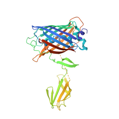
7Y96 | pdb_00007y96

Crystal structure of the carboxy-terminal domain of a coronavirus M protein fused with a split GFP


Experimental Data Snapshot

  • Method: X-RAY DIFFRACTION
  • Resolution: 3.42 Å
  • R-Value Free: 
    0.265 (Depositor), 0.270 (DCC) 
  • R-Value Work: 
    0.240 (Depositor), 0.240 (DCC) 
  • R-Value Observed: 
    0.241 (Depositor) 

Starting Model: experimental
View more details

wwPDB Validation   3D Report Full Report


This is version 1.5 of the entry. See complete history


Literature

Crystal structure of the membrane (M) protein from a bat betacoronavirus.

Wang, X.Yang, Y.Sun, Z.Zhou, X.

(2023) PNAS Nexus 2: pgad021-pgad021

  • DOI: https://doi.org/10.1093/pnasnexus/pgad021
  • Primary Citation of Related Structures:  
    7Y96, 7Y9B

  • PubMed Abstract: 

    The membrane (M) protein is the most abundant structural protein of coronaviruses including MERS-CoV, SARS-CoV, and SARS-CoV-2, and plays a central role in virus assembly through its interaction with various partner proteins. However, mechanistic details about how M protein interacts with others remain elusive due to lack of high-resolution structures. Here, we present the first crystal structure of a betacoronavirus M protein from Pipistrellus bat coronavirus HKU5 (batCOV5-M), which is closely related to MERS-CoV, SARS-CoV, and SARS-CoV-2 M proteins. Furthermore, an interaction analysis indicates that the carboxy-terminus of the batCOV5 nucleocapsid (N) protein mediates its interaction with batCOV5-M. Combined with a computational docking analysis an M-N interaction model is proposed, providing insight into the mechanism of M protein-mediated protein interactions.


  • Organizational Affiliation
    • State Key Laboratory of Biotherapy, Department of Integrated Traditional Chinese and Western Medicine, Rare Diseases Center, West China Hospital, Sichuan University, Chengdu, Sichuan 610041, China.

Macromolecules
Find similar proteins by:  (by identity cutoff)  |  3D Structure
Entity ID: 1
MoleculeChains Sequence LengthOrganismDetailsImage
Green fluorescent protein,Membrane protein
A, B
340Aequorea victoriaPipistrellus bat coronavirus HKU5Mutation(s): 11 
Gene Names: GFPM5
UniProt
Find proteins for A3EXD6 (Bat coronavirus HKU5)
Explore A3EXD6 
Go to UniProtKB:  A3EXD6
Find proteins for P42212 (Aequorea victoria)
Explore P42212 
Go to UniProtKB:  P42212
Entity Groups  
Sequence Clusters30% Identity50% Identity70% Identity90% Identity95% Identity100% Identity
UniProt GroupsP42212A3EXD6
Sequence Annotations
Expand
  • Reference Sequence
Small Molecules
Modified Residues  1 Unique
IDChains TypeFormula2D DiagramParent
CRO
Query on CRO
A, B
L-PEPTIDE LINKINGC15 H17 N3 O5THR, TYR, GLY
Experimental Data & Validation

Experimental Data

  • Method: X-RAY DIFFRACTION
  • Resolution: 3.42 Å
  • R-Value Free:  0.265 (Depositor), 0.270 (DCC) 
  • R-Value Work:  0.240 (Depositor), 0.240 (DCC) 
  • R-Value Observed: 0.241 (Depositor) 
Space Group: P 61 2 2
Unit Cell:
Length ( Å )Angle ( ˚ )
a = 82.02α = 90
b = 82.02β = 90
c = 427.841γ = 120
Software Package:
Software NamePurpose
PHENIXrefinement
XDSdata reduction
Aimlessdata scaling
PHASERphasing
PDB_EXTRACTdata extraction

Structure Validation

View Full Validation Report


Entry History & Funding Information

Deposition Data


Funding OrganizationLocationGrant Number
National Natural Science Foundation of China (NSFC)China31770783

Revision History  (Full details and data files)

  • Version 1.0: 2022-08-17
    Type: Initial release
  • Version 1.1: 2023-03-01
    Changes: Database references
  • Version 1.2: 2023-03-22
    Changes: Database references
  • Version 1.3: 2023-05-31
    Changes: Database references, Refinement description
  • Version 1.4: 2023-11-29
    Changes: Data collection, Refinement description
  • Version 1.5: 2024-11-06
    Changes: Structure summary