8QM5 | pdb_00008qm5

Potential drug binding sites for translation initiation factor eIF4E


Experimental Data Snapshot

  • Method: X-RAY DIFFRACTION
  • Resolution: 1.89 Å
  • R-Value Free: 
    0.220 (Depositor), 0.219 (DCC) 
  • R-Value Work: 
    0.194 (Depositor), 0.198 (DCC) 
  • R-Value Observed: 
    0.196 (Depositor) 

wwPDB Validation   3D Report Full Report


Ligand Structure Quality Assessment 


This is version 1.1 of the entry. See complete history


Literature

Integrating fragment-based screening with targeted protein degradation and genetic rescue to explore eIF4E function.

Sharp, S.Y.Martella, M.D'Agostino, S.Milton, C.I.Ward, G.Woodhead, A.J.Richardson, C.J.Carr, M.G.Chiarparin, E.Cons, B.D.Coyle, J.East, C.E.Hiscock, S.D.Martinez-Fleites, C.Mortenson, P.N.Palmer, N.Pathuri, P.Powers, M.V.Saalau, S.M.St Denis, J.D.Swabey, K.Vinkovic, M.Walton, H.Williams, G.Clarke, P.A.

(2024) Nat Commun 15: 10037-10037

  • DOI: https://doi.org/10.1038/s41467-024-54356-1
  • Primary Citation of Related Structures:  
    8QM4, 8QM5, 8QM6, 8QM7, 8QM8, 8QM9

  • PubMed Abstract: 

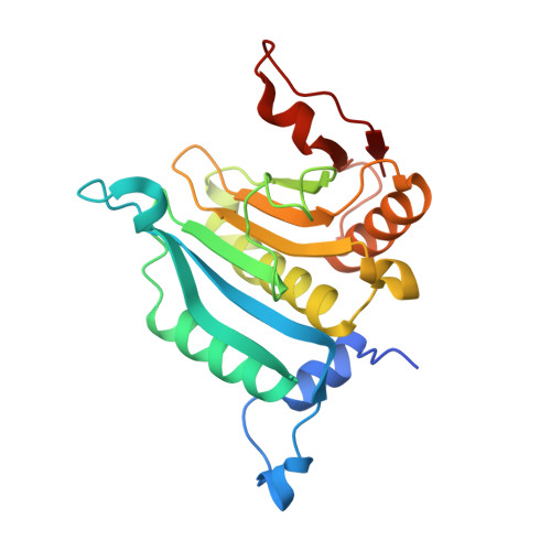
    Eukaryotic initiation factor 4E (eIF4E) serves as a regulatory hub for oncogene-driven protein synthesis and is considered a promising anticancer target. Here we screen a fragment library against eIF4E and identify a ligand-binding site with previously unknown function. Follow-up structure-based design yields a low nM tool compound (4, K d  = 0.09 µM; LE 0.38), which disrupts the eIF4E:eIF4G interaction, inhibits translation in cell lysates, and demonstrates target engagement with eIF4E in intact cells (EC 50  = 2 µM). By coupling targeted protein degradation with genetic rescue using eIF4E mutants, we show that disruption of both the canonical eIF4G and non-canonical binding sites is likely required to drive a strong cellular effect. This work highlights the power of fragment-based drug discovery to identify pockets in difficult-to-drug proteins and how this approach can be combined with genetic characterization and degrader technology to probe protein function in complex biological systems.


  • Organizational Affiliation

    RNA Biology and Molecular Therapeutics Team, Centre for Cancer Drug Discovery, Institute of Cancer Research, London, SM2 5NG, UK.


Macromolecules
Find similar proteins by:  (by identity cutoff)  |  3D Structure
Entity ID: 1
MoleculeChains Sequence LengthOrganismDetailsImage
Eukaryotic translation initiation factor 4E
A, B
215Homo sapiensMutation(s): 0 
Gene Names: EIF4E
UniProt & NIH Common Fund Data Resources
Find proteins for P06730 (Homo sapiens)
Explore P06730 
Go to UniProtKB:  P06730
PHAROS:  P06730
GTEx:  ENSG00000151247 
Entity Groups  
Sequence Clusters30% Identity50% Identity70% Identity90% Identity95% Identity100% Identity
UniProt GroupP06730
Sequence Annotations
Expand
  • Reference Sequence
Experimental Data & Validation

Experimental Data

  • Method: X-RAY DIFFRACTION
  • Resolution: 1.89 Å
  • R-Value Free:  0.220 (Depositor), 0.219 (DCC) 
  • R-Value Work:  0.194 (Depositor), 0.198 (DCC) 
  • R-Value Observed: 0.196 (Depositor) 
Space Group: P 1 21 1
Unit Cell:
Length ( Å )Angle ( ˚ )
a = 47.124α = 90
b = 68.877β = 96.57
c = 63.729γ = 90
Software Package:
Software NamePurpose
BUSTERrefinement
XDSdata reduction
SCALAdata scaling
BUSTERphasing

Structure Validation

View Full Validation Report


Ligand Structure Quality Assessment 


Entry History & Funding Information

Deposition Data

  • Released Date: 2025-01-15 
  • Deposition Author(s): Cleasby, A.

Funding OrganizationLocationGrant Number
Not funded--

Revision History  (Full details and data files)

  • Version 1.0: 2025-01-15
    Type: Initial release
  • Version 1.1: 2025-03-12
    Changes: Database references