8U1I | pdb_00008u1i

Crystal structure of SyoA bound to 4-methylsyringol


Experimental Data Snapshot

  • Method: X-RAY DIFFRACTION
  • Resolution: 1.12 Å
  • R-Value Free: 
    0.153 (Depositor), 0.150 (DCC) 
  • R-Value Work: 
    0.137 (Depositor), 0.140 (DCC) 
  • R-Value Observed: 
    0.138 (Depositor) 

Starting Model: experimental
View more details

wwPDB Validation   3D Report Full Report


Ligand Structure Quality Assessment 


This is version 1.2 of the entry. See complete history


Literature

Structural insights into S-lignin O-demethylation via a rare class of heme peroxygenase enzymes.

Harlington, A.C.Das, T.Shearwin, K.E.Bell, S.G.Whelan, F.

(2025) Nat Commun 16: 1815-1815

  • DOI: https://doi.org/10.1038/s41467-025-57129-6
  • Primary Citation of Related Structures:  
    8U09, 8U19, 8U1I

  • PubMed Abstract: 

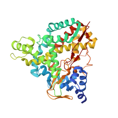
    The O-demethylation of lignin aromatics is a rate-limiting step in their bioconversion to higher-value compounds. A recently discovered cytochrome P450, SyoA, demethylates the S-lignin aromatic syringol. In this work, we solve high-resolution X-ray crystal structures of substrate-free and substrate-bound SyoA and evaluate demethylation of para-substituted S-lignin aromatics via monooxygenase and peroxide shunt pathways. We find that SyoA demethylates S-lignin aromatics exclusively using the peroxide shunt pathway. The atomic-resolution structures reveal the position of non-canonical residues in the I-helix (Gln252, Glu253). Mutagenesis of this amide-acid pair in SyoA shows they are critical for catalytic activity. This work expands the enzymatic toolkit for improving the capacity to funnel lignin derived aromatics towards higher value compounds and defines the chemistry within the active site of the enzyme that enables peroxygenase activity. These insights provide a framework for engineering peroxygenase activity in other heme enzymes to generate easier to use biocatalysts.


  • Organizational Affiliation

    Department of Molecular and Biomedical Science, University of Adelaide, Adelaide, SA, Australia.


Macromolecules
Find similar proteins by:  (by identity cutoff)  |  3D Structure
Entity ID: 1
MoleculeChains Sequence LengthOrganismDetailsImage
Cytochrome P450412Amycolatopsis thermoflava N1165Mutation(s): 0 
EC: 1.14.14.1
UniProt
Find proteins for A0AAX7FLX8 (Amycolatopsis thermoflava N1165)
Explore A0AAX7FLX8 
Go to UniProtKB:  A0AAX7FLX8
Entity Groups  
Sequence Clusters30% Identity50% Identity70% Identity90% Identity95% Identity100% Identity
UniProt GroupA0AAX7FLX8
Sequence Annotations
Expand
  • Reference Sequence
Experimental Data & Validation

Experimental Data

  • Method: X-RAY DIFFRACTION
  • Resolution: 1.12 Å
  • R-Value Free:  0.153 (Depositor), 0.150 (DCC) 
  • R-Value Work:  0.137 (Depositor), 0.140 (DCC) 
  • R-Value Observed: 0.138 (Depositor) 
Space Group: P 1 21 1
Unit Cell:
Length ( Å )Angle ( ˚ )
a = 57.414α = 90
b = 62.764β = 104.58
c = 58.078γ = 90
Software Package:
Software NamePurpose
PHENIXrefinement
XDSdata reduction
Aimlessdata scaling
PHENIXphasing

Structure Validation

View Full Validation Report


Ligand Structure Quality Assessment 


Entry History & Funding Information

Deposition Data


Funding OrganizationLocationGrant Number
Wine AustraliaAustraliaPH2002
Australian Research Council (ARC)AustraliaDP200102411
Australian Research Council (ARC)AustraliaDP230103062

Revision History  (Full details and data files)

  • Version 1.0: 2024-07-24
    Type: Initial release
  • Version 1.1: 2025-03-05
    Changes: Database references, Structure summary
  • Version 1.2: 2025-03-19
    Changes: Author supporting evidence