8WLW | pdb_00008wlw

Crystal structure of DelP123_Msd in complex with 5-azauracil


Experimental Data Snapshot

  • Method: X-RAY DIFFRACTION
  • Resolution: 1.54 Å
  • R-Value Free: 
    0.209 (Depositor), 0.222 (DCC) 
  • R-Value Work: 
    0.176 (Depositor), 0.191 (DCC) 

Starting Model: experimental
View more details

wwPDB Validation   3D Report Full Report


Ligand Structure Quality Assessment 


This is version 1.2 of the entry. See complete history


Literature

Mechanism of deamination by mycobacterial deaminase selectively targeting mutagenic bases.

Porathoor, S.Choudhury, A.R.Chakrabarti, R.Anand, R.

(2025) Nucleic Acids Res 53

  • DOI: https://doi.org/10.1093/nar/gkaf171
  • Primary Citation of Related Structures:  
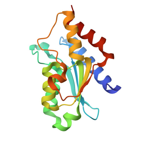
    8WLV, 8WLW, 8WLX

  • PubMed Abstract: 

    Nucleobase deaminases are important players in maintaining a stringent nucleobase pool and enhancing genetic diversity via judicious base editing. Here, we delineate the mechanism of Mycobacterium smegmatis deaminase, Msd, found predominantly in Mycobacterium species, that selectively catalyzes the deamination of mutagenic bases. Molecular dynamic studies reveal the dynamic nature of unique structural insertions that cycle between 'closed' and 'open' states, enabling zinc-assisted deamination by occluding the solvent. Corroborating X-ray crystallographic and biochemical studies assert that the appropriate length of the two gating loops and proper positioning of the di-proline motif they harbor are paramount to effective closure. Analysis reveals that although both natural base deaminases, guanine and cytosine deaminase, operate via a similar gating mechanism to Msd, they employ topologically differentially placed structural elements to achieve the 'closed' form. The comparison shows that Mycobacterium deaminases lack the dual-proton shuttle, which renders them ineffective for the deamination of natural bases but allows them to selectively target mutagenic s-triazine scaffolds, thereby imparting innate immunity against these drugs. The study highlights how topologically unique insertions in the cytidine deaminase fold play a critical role in harnessing evolutionary versatility, responsible in imparting fidelity for a nucleobase deamination reaction.


  • Organizational Affiliation

    Department of Chemistry, Indian Institute of Technology Bombay, Mumbai, Maharashtra 400076, India.


Macromolecules
Find similar proteins by:  (by identity cutoff)  |  3D Structure
Entity ID: 1
MoleculeChains Sequence LengthOrganismDetailsImage
CMP/dCMP deaminase, zinc-binding proteinA,
B [auth C],
C [auth B],
D
157Mycolicibacterium smegmatis MC2 155Mutation(s): 0 
Gene Names: MSMEI_3493
UniProt
Find proteins for A0QY90 (Mycolicibacterium smegmatis (strain ATCC 700084 / mc(2)155))
Explore A0QY90 
Go to UniProtKB:  A0QY90
Entity Groups  
Sequence Clusters30% Identity50% Identity70% Identity90% Identity95% Identity100% Identity
UniProt GroupA0QY90
Sequence Annotations
Expand
  • Reference Sequence
Small Molecules
Ligands 3 Unique
IDChains Name / Formula / InChI Key2D Diagram3D Interactions
WHC (Subject of Investigation/LOI)
Query on WHC

Download Ideal Coordinates CCD File 
E [auth A],
H [auth C],
L [auth B],
O [auth D]
1~{H}-1,3,5-triazine-2,4-dione
C3 H3 N3 O2
GEWRKGDRYZIFNP-UHFFFAOYSA-N
GOL
Query on GOL

Download Ideal Coordinates CCD File 
F [auth A],
I [auth C],
J [auth C],
M [auth B]
GLYCEROL
C3 H8 O3
PEDCQBHIVMGVHV-UHFFFAOYSA-N
ZN (Subject of Investigation/LOI)
Query on ZN

Download Ideal Coordinates CCD File 
G [auth A],
K [auth C],
N [auth B],
P [auth D]
ZINC ION
Zn
PTFCDOFLOPIGGS-UHFFFAOYSA-N
Experimental Data & Validation

Experimental Data

  • Method: X-RAY DIFFRACTION
  • Resolution: 1.54 Å
  • R-Value Free:  0.209 (Depositor), 0.222 (DCC) 
  • R-Value Work:  0.176 (Depositor), 0.191 (DCC) 
Space Group: P 1 21 1
Unit Cell:
Length ( Å )Angle ( ˚ )
a = 46.75α = 90
b = 85.3β = 102.491
c = 76.46γ = 90
Software Package:
Software NamePurpose
REFMACrefinement
XDSdata reduction
XSCALEdata scaling
PHASERphasing

Structure Validation

View Full Validation Report


Ligand Structure Quality Assessment 


Entry History & Funding Information

Deposition Data


Funding OrganizationLocationGrant Number
Department of Biotechnology (DBT, India)IndiaBT/HRD/NWBA/39/03/2018-19

Revision History  (Full details and data files)

  • Version 1.0: 2024-09-18
    Type: Initial release
  • Version 1.1: 2025-04-02
    Changes: Database references, Structure summary
  • Version 1.2: 2025-04-16
    Changes: Database references