8C61 | pdb_00008c61

Structure of USP54 in complex with Lys63-linked diUbiquitin-PA


Experimental Data Snapshot

  • Method: X-RAY DIFFRACTION
  • Resolution: 2.50 Å
  • R-Value Free: 
    0.243 (Depositor), 0.245 (DCC) 
  • R-Value Work: 
    0.200 (Depositor), 0.203 (DCC) 
  • R-Value Observed: 
    0.203 (Depositor) 

Starting Models: experimental, in silico
View more details

wwPDB Validation   3D Report Full Report


Ligand Structure Quality Assessment 


This is version 1.1 of the entry. See complete history


Literature

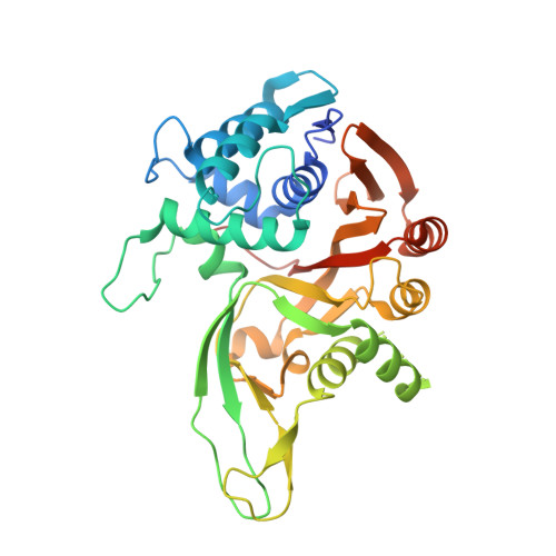
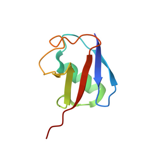
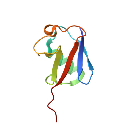
Discovery and mechanism of K63-linkage-directed deubiquitinase activity in USP53.

Wendrich, K.Gallant, K.Recknagel, S.Petroulia, S.Kazi, N.H.Hane, J.A.Fuhrer, S.Bezstarosti, K.O'Dea, R.Demmers, J.Gersch, M.

(2024) Nat Chem Biol 

  • DOI: https://doi.org/10.1038/s41589-024-01777-0
  • Primary Citation of Related Structures:  
    8C61

  • PubMed Abstract: 

    Ubiquitin-specific proteases (USPs) represent the largest class of human deubiquitinases (DUBs) and comprise its phylogenetically most distant members USP53 and USP54, which are annotated as catalytically inactive pseudoenzymes. Conspicuously, mutations within the USP domain of USP53 cause progressive familial intrahepatic cholestasis. Here, we report the discovery that USP53 and USP54 are active DUBs with high specificity for K63-linked polyubiquitin. We demonstrate how USP53 mutations abrogate catalytic activity, implicating loss of DUB activity in USP53-mediated pathology. Depletion of USP53 increases K63-linked ubiquitination of tricellular junction components. Assays with substrate-bound polyubiquitin reveal that USP54 cleaves within K63-linked chains, whereas USP53 can en bloc deubiquitinate substrate proteins in a K63-linkage-dependent manner. Biochemical and structural analyses uncover underlying K63-specific S2 ubiquitin-binding sites within their catalytic domains. Collectively, our work revises the annotation of USP53 and USP54, provides reagents and a mechanistic framework to investigate K63-linked polyubiquitin decoding and establishes K63-linkage-directed deubiquitination as a new DUB activity.


  • Organizational Affiliation

    Chemical Genomics Centre, Max Planck Institute of Molecular Physiology, Dortmund, Germany.


Macromolecules
Find similar proteins by:  (by identity cutoff)  |  3D Structure
Entity ID: 1
MoleculeChains Sequence LengthOrganismDetailsImage
Inactive ubiquitin carboxyl-terminal hydrolase 54
A, D, G, J
350Homo sapiensMutation(s): 0 
Gene Names: USP54C10orf29
EC: 3.4.19.12
UniProt & NIH Common Fund Data Resources
Find proteins for Q70EL1 (Homo sapiens)
Explore Q70EL1 
Go to UniProtKB:  Q70EL1
PHAROS:  Q70EL1
GTEx:  ENSG00000166348 
Entity Groups  
Sequence Clusters30% Identity50% Identity70% Identity90% Identity95% Identity100% Identity
UniProt GroupQ70EL1
Sequence Annotations
Expand
  • Reference Sequence
Find similar proteins by:  (by identity cutoff)  |  3D Structure
Entity ID: 2
MoleculeChains Sequence LengthOrganismDetailsImage
Polyubiquitin-B
B, E, H, K
75Homo sapiensMutation(s): 0 
Gene Names: UBB
UniProt & NIH Common Fund Data Resources
Find proteins for P0CG47 (Homo sapiens)
Explore P0CG47 
Go to UniProtKB:  P0CG47
PHAROS:  P0CG47
GTEx:  ENSG00000170315 
Entity Groups  
Sequence Clusters30% Identity50% Identity70% Identity90% Identity95% Identity100% Identity
UniProt GroupP0CG47
Sequence Annotations
Expand
  • Reference Sequence
Find similar proteins by:  (by identity cutoff)  |  3D Structure
Entity ID: 3
MoleculeChains Sequence LengthOrganismDetailsImage
Ubiquitin
C, F, I, L
76Homo sapiensMutation(s): 1 
Gene Names: UBB
UniProt & NIH Common Fund Data Resources
Find proteins for P0CG47 (Homo sapiens)
Explore P0CG47 
Go to UniProtKB:  P0CG47
PHAROS:  P0CG47
GTEx:  ENSG00000170315 
Entity Groups  
Sequence Clusters30% Identity50% Identity70% Identity90% Identity95% Identity100% Identity
UniProt GroupP0CG47
Sequence Annotations
Expand
  • Reference Sequence
Small Molecules
Ligands 4 Unique
IDChains Name / Formula / InChI Key2D Diagram3D Interactions
ZN (Subject of Investigation/LOI)
Query on ZN

Download Ideal Coordinates CCD File 
CA [auth J]
DA [auth J]
EA [auth J]
M [auth A]
N [auth A]
CA [auth J],
DA [auth J],
EA [auth J],
M [auth A],
N [auth A],
O [auth A],
T [auth D],
U [auth D],
V [auth D],
X [auth G],
Y [auth G],
Z [auth G]
ZINC ION
Zn
PTFCDOFLOPIGGS-UHFFFAOYSA-N
AYE (Subject of Investigation/LOI)
Query on AYE

Download Ideal Coordinates CCD File 
AA [auth G],
FA [auth J],
P [auth A],
W [auth D]
prop-2-en-1-amine
C3 H7 N
VVJKKWFAADXIJK-UHFFFAOYSA-N
CL
Query on CL

Download Ideal Coordinates CCD File 
Q [auth A],
R [auth A]
CHLORIDE ION
Cl
VEXZGXHMUGYJMC-UHFFFAOYSA-M
NA
Query on NA

Download Ideal Coordinates CCD File 
BA [auth G],
S [auth C]
SODIUM ION
Na
FKNQFGJONOIPTF-UHFFFAOYSA-N
Experimental Data & Validation

Experimental Data

  • Method: X-RAY DIFFRACTION
  • Resolution: 2.50 Å
  • R-Value Free:  0.243 (Depositor), 0.245 (DCC) 
  • R-Value Work:  0.200 (Depositor), 0.203 (DCC) 
  • R-Value Observed: 0.203 (Depositor) 
Space Group: P 21 21 21
Unit Cell:
Length ( Å )Angle ( ˚ )
a = 122.743α = 90
b = 126.559β = 90
c = 144.167γ = 90
Software Package:
Software NamePurpose
PHENIXrefinement
DIALSdata reduction
Aimlessdata scaling
CRANK2phasing
Cootmodel building
PHASERphasing

Structure Validation

View Full Validation Report


Ligand Structure Quality Assessment 


Entry History & Funding Information

Deposition Data


Funding OrganizationLocationGrant Number
Max Planck SocietyGermanyCGC-III-352S

Revision History  (Full details and data files)

  • Version 1.0: 2024-07-31
    Type: Initial release
  • Version 1.1: 2025-02-12
    Changes: Database references, Derived calculations, Structure summary