


Structure of the LpMIP mutant D44E
Wiedemann, C., Whittaker, J.J., Guskov, A., Hellmich, U.A.To be published.
Experimental Data Snapshot
Starting Model: experimental
View more details
wwPDB Validation 3D Report Full Report

Entity ID: 1 | |||||
|---|---|---|---|---|---|
| Molecule | Chains | Sequence Length | Organism | Details | Image |
| Outer membrane protein MIP | 205 | Legionella pneumophila | Mutation(s): 1 Gene Names: mip EC: 5.2.1.8 | 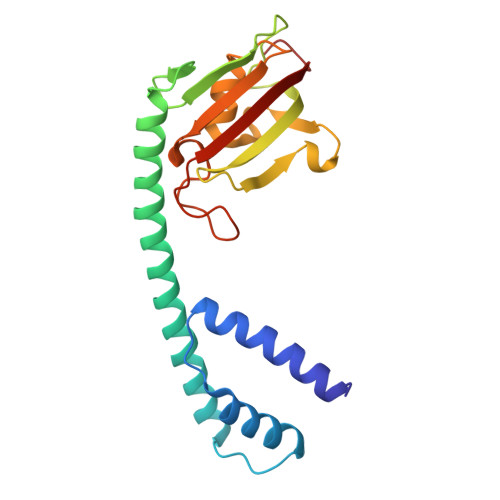 | |
UniProt | |||||
Find proteins for Q70YI1 (Legionella pneumophila) Explore Q70YI1 Go to UniProtKB: Q70YI1 | |||||
Entity Groups | |||||
| Sequence Clusters | 30% Identity50% Identity70% Identity90% Identity95% Identity100% Identity | ||||
| UniProt Group | Q70YI1 | ||||
Sequence AnnotationsExpand | |||||
| |||||
| Ligands 2 Unique | |||||
|---|---|---|---|---|---|
| ID | Chains | Name / Formula / InChI Key | 2D Diagram | 3D Interactions | |
| MES Query on MES | B [auth A] | 2-(N-MORPHOLINO)-ETHANESULFONIC ACID C6 H13 N O4 S SXGZJKUKBWWHRA-UHFFFAOYSA-N |  | ||
| PEG Query on PEG | C [auth A], D [auth A], E [auth A] | DI(HYDROXYETHYL)ETHER C4 H10 O3 MTHSVFCYNBDYFN-UHFFFAOYSA-N |  | ||
| Length ( Å ) | Angle ( ˚ ) |
|---|---|
| a = 77.185 | α = 90 |
| b = 77.185 | β = 90 |
| c = 103.517 | γ = 90 |
| Software Name | Purpose |
|---|---|
| REFMAC | refinement |
| XDS | data scaling |
| XDS | data reduction |
| MoRDa | phasing |
| Funding Organization | Location | Grant Number |
|---|---|---|
| German Research Foundation (DFG) | Germany | 390713860 |


