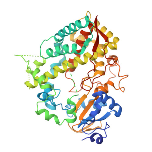
9COS | pdb_00009cos

Crystal structure of CYP3A4 bound to an inhibitor


Experimental Data Snapshot

  • Method: X-RAY DIFFRACTION
  • Resolution: 2.75 Å
  • R-Value Free: 
    0.280 (Depositor), 0.288 (DCC) 
  • R-Value Work: 
    0.244 (Depositor), 0.249 (DCC) 
  • R-Value Observed: 
    0.247 (Depositor) 

Starting Model: experimental
View more details

wwPDB Validation   3D Report Full Report


Ligand Structure Quality Assessment 


This is version 1.1 of the entry. See complete history


Literature

Evaluation of Larger Side-Group Functionalities and the Side/End-Group Interplay in Ritonavir-Like Inhibitors of CYP3A4.

Samuels, E.R.Sevrioukova, I.F.

(2025) Chem Biol Drug Des 105: e70043-e70043

  • DOI: https://doi.org/10.1111/cbdd.70043
  • Primary Citation of Related Structures:  
    9COR, 9COS, 9COT, 9COU, 9COV, 9COW, 9COX, 9COY

  • PubMed Abstract: 

    A new series of 13 ritonavir-like inhibitors of human drug-metabolizing CYP3A4 was rationally designed to study the R 2 side-group and R 3 end-group interplay when the R 1 side-group is represented by phenyl. Spectral, functional, and structural characterization showed no improvement in the binding affinity and inhibitory potency of R 1 /R 2 -phenyl inhibitors upon elongation and/or fluorination of R 3 -Boc (tert-butyloxycarbonyl) or its replacement with benzenesulfonyl. When R 3 is pyridine, the impact of R 2 -phenyl-to-indole/naphthalene substitution was multidirectional and highly dependent on side-group stereo configuration. Overall, the R 2 -naphthalene/R 3 -pyridine containing 2f (R/S) was the series lead compound and one of the strongest binders/inhibitors designed thus far (K s  = 0.009 μM; IC 50  = 0.10 μM). Introduction of a larger biphenyl or fluorene as R 2 did not lead to any improvements. Contrarily, fluorene-containing 13 was the series weakest binder and inhibitor (K s  = 0.734 μM; IC 50  = 1.32 μM), implying that the fluorene moiety is too large to allow unrestricted access to the active site. The R 2 -biphenyl, however, can switch positions with R 3 -Boc to enable heme ligation. Thus, for small and chemically simple end-groups such as Boc and pyridine, the R 2 /R 3 interplay could lead to conformational rearrangement that would be difficult to foresee without structural information.


  • Organizational Affiliation

    Department of Pharmaceutical Sciences, University of California, Irvine, California, USA.


Macromolecules
Find similar proteins by:  (by identity cutoff)  |  3D Structure
Entity ID: 1
MoleculeChains Sequence LengthOrganismDetailsImage
Cytochrome P450 3A4487Homo sapiensMutation(s): 0 
Gene Names: CYP3A4CYP3A3
EC: 1.14.14.1 (PDB Primary Data), 1.14.14.56 (PDB Primary Data), 1.14.14.73 (PDB Primary Data), 1.14.14.55 (PDB Primary Data)
UniProt & NIH Common Fund Data Resources
Find proteins for P08684 (Homo sapiens)
Explore P08684 
Go to UniProtKB:  P08684
PHAROS:  P08684
GTEx:  ENSG00000160868 
Entity Groups  
Sequence Clusters30% Identity50% Identity70% Identity90% Identity95% Identity100% Identity
UniProt GroupP08684
Sequence Annotations
Expand
  • Reference Sequence
Small Molecules
Ligands 2 Unique
IDChains Name / Formula / InChI Key2D Diagram3D Interactions
HEM
Query on HEM

Download Ideal Coordinates CCD File 
B [auth A]PROTOPORPHYRIN IX CONTAINING FE
C34 H32 Fe N4 O4
KABFMIBPWCXCRK-RGGAHWMASA-L
A1A06 (Subject of Investigation/LOI)
Query on A1A06

Download Ideal Coordinates CCD File 
C [auth A](2R)-3-phenyl-2-{[(2S)-3-phenyl-2-{[N-(trifluoroacetyl)glycyl]amino}propyl]sulfanyl}-N-[3-(pyridin-3-yl)propyl]propanamide
C30 H33 F3 N4 O3 S
DWYGERMVFBUIQJ-IZZNHLLZSA-N
Experimental Data & Validation

Experimental Data

  • Method: X-RAY DIFFRACTION
  • Resolution: 2.75 Å
  • R-Value Free:  0.280 (Depositor), 0.288 (DCC) 
  • R-Value Work:  0.244 (Depositor), 0.249 (DCC) 
  • R-Value Observed: 0.247 (Depositor) 
Space Group: I 2 2 2
Unit Cell:
Length ( Å )Angle ( ˚ )
a = 76.19α = 90
b = 100.16β = 90
c = 124.73γ = 90
Software Package:
Software NamePurpose
PHENIXrefinement
MOSFLMdata reduction
SCALAdata scaling
PHASERphasing

Structure Validation

View Full Validation Report


Ligand Structure Quality Assessment 


Entry History & Funding Information

Deposition Data


Funding OrganizationLocationGrant Number
National Institutes of Health/National Institute of Environmental Health Sciences (NIH/NIEHS)United StatesES025767

Revision History  (Full details and data files)

  • Version 1.0: 2025-01-08
    Type: Initial release
  • Version 1.1: 2025-03-05
    Changes: Database references