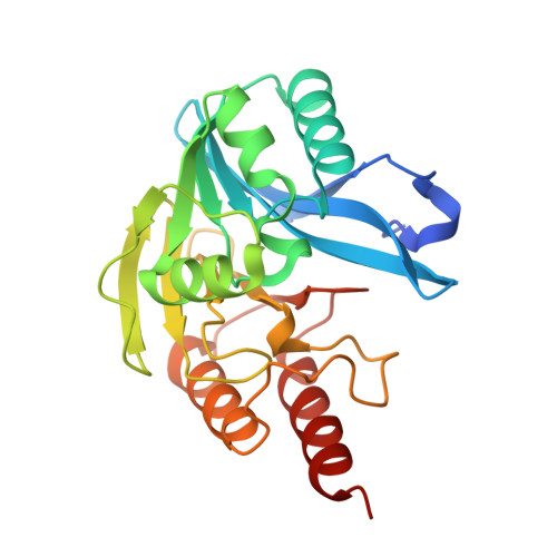
9CV3 | pdb_00009cv3

Crystal structure of the metallo-beta-lactamase VIM-20 with L-captopril


Experimental Data Snapshot

  • Method: X-RAY DIFFRACTION
  • Resolution: 1.51 Å
  • R-Value Free: 
    0.212 (Depositor), 0.212 (DCC) 
  • R-Value Work: 
    0.192 (Depositor), 0.192 (DCC) 
  • R-Value Observed: 
    0.193 (Depositor) 

Starting Model: experimental
View more details

wwPDB Validation   3D Report Full Report


Ligand Structure Quality Assessment 


This is version 1.0 of the entry. See complete history


Literature

Mutation of an active site-adjacent residue in VIM indirectly dictates interactions with and blunts inhibition by D-captopril.

Silwal, S.B.Wamsley, B.Wang, Z.Gung, B.W.Nix, J.C.Page, R.C.

(2025) J Inorg Biochem 271: 112975-112975

  • DOI: https://doi.org/10.1016/j.jinorgbio.2025.112975
  • Primary Citation of Related Structures:  
    9CV1, 9CV2, 9CV3, 9CV4, 9CV5

  • PubMed Abstract: 

    Activity assays and X-ray crystallographic studies were undertaken to elucidate the inhibitory mechanism of captopril stereoisomers on Verona integron-encoded metallo-β-lactamases, specifically VIM-20, VIM-31, and VIM-15. All three VIM-2-like variants (VIM-20, VIM-31, and VIM-15) and VIM-2 expressed in Escherichia coli exhibited catalytic activity with comparable steady-state kinetic parameters. Among the tested thiol drugs (L- and D-captopril, D,L-thiorphan, and 2,3-dimercaprol), IC 50 analyses indicated that D-captopril and 2,3-dimercaprol were more potent inhibitors against the VIM enzymes examined in this study. Notably, the IC 50 value of D-captopril against VIM-31 was an exception, closely resembling that of L-captopril. To elucidate this exceptional inhibitory potency of D-captopril and its binding mode in the active site of VIM-31, high-resolution crystal structures of VIM-20, VIM-31, and VIM-15 in complex with both L- and D-captopril are reported. These findings will help evaluate whether the identified potent inhibitor D-captopril could be further developed as a pan inhibitor targeting the VIM-family enzymes.


  • Organizational Affiliation

    Department of Chemistry and Biochemistry, Miami University, Oxford, OH, USA.


Macromolecules
Find similar proteins by:  (by identity cutoff)  |  3D Structure
Entity ID: 1
MoleculeChains Sequence LengthOrganismDetailsImage
Metallo-beta-lactamase VIM-20243Enterobacter cloacaeMutation(s): 0 
Gene Names: blaVIM-20
EC: 3.5.2.6
UniProt
Find proteins for A0A344X7M2 (Enterobacter cloacae)
Explore A0A344X7M2 
Go to UniProtKB:  A0A344X7M2
Entity Groups  
Sequence Clusters30% Identity50% Identity70% Identity90% Identity95% Identity100% Identity
UniProt GroupA0A344X7M2
Sequence Annotations
Expand
  • Reference Sequence
Small Molecules
Ligands 3 Unique
IDChains Name / Formula / InChI Key2D Diagram3D Interactions
X8Z (Subject of Investigation/LOI)
Query on X8Z

Download Ideal Coordinates CCD File 
E [auth A]L-CAPTOPRIL
C9 H15 N O3 S
FAKRSMQSSFJEIM-RQJHMYQMSA-N
ZN
Query on ZN

Download Ideal Coordinates CCD File 
B [auth A],
C [auth A],
D [auth A]
ZINC ION
Zn
PTFCDOFLOPIGGS-UHFFFAOYSA-N
ACT
Query on ACT

Download Ideal Coordinates CCD File 
F [auth A],
G [auth A],
H [auth A]
ACETATE ION
C2 H3 O2
QTBSBXVTEAMEQO-UHFFFAOYSA-M
Experimental Data & Validation

Experimental Data

  • Method: X-RAY DIFFRACTION
  • Resolution: 1.51 Å
  • R-Value Free:  0.212 (Depositor), 0.212 (DCC) 
  • R-Value Work:  0.192 (Depositor), 0.192 (DCC) 
  • R-Value Observed: 0.193 (Depositor) 
Space Group: I 2 2 2
Unit Cell:
Length ( Å )Angle ( ˚ )
a = 67.845α = 90
b = 77.595β = 90
c = 79.052γ = 90
Software Package:
Software NamePurpose
PHENIXrefinement
XDSdata reduction
XDSdata scaling
PHASERphasing

Structure Validation

View Full Validation Report


Ligand Structure Quality Assessment 


Entry History & Funding Information

Deposition Data


Funding OrganizationLocationGrant Number
National Institutes of Health/National Institute of General Medical Sciences (NIH/NIGMS)United States128595

Revision History  (Full details and data files)

  • Version 1.0: 2025-07-09
    Type: Initial release