Conformational flexibility is a critical factor in designing broad-spectrum human norovirus protease inhibitors.
Pham, S., Zhao, B., Neetu, N., Sankaran, B., Patil, K., Ramani, S., Song, Y., Estes, M.K., Palzkill, T., Prasad, B.V.V.(2025) J Virol 99: e0175724-e0175724
- PubMed: 39873493
- DOI: https://doi.org/10.1128/jvi.01757-24
- Primary Citation of Related Structures:
9D9T, 9D9Y, 9DA0, 9DA7, 9DAJ, 9DAL, 9DAP, 9DEY, 9DF5 - PubMed Abstract:
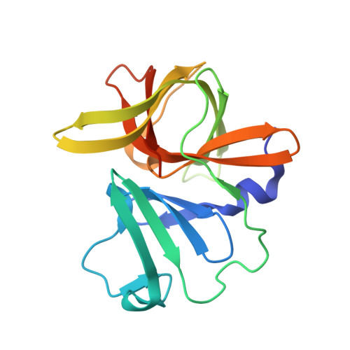
Human norovirus (HuNoV) is a leading cause of gastroenteritis worldwide and is associated with significant morbidity, mortality, and economic impact. There are currently no licensed antiviral drugs for the treatment of HuNoV-associated gastroenteritis. The HuNoV protease plays a critical role in the initiation of virus replication by cleaving the polyprotein. Thus, it is an ideal target for developing antiviral small-molecule inhibitors. While rupintrivir, a potent small-molecule inhibitor of several picornavirus proteases, effectively inhibits GI.1 protease, it is an order of magnitude less effective against GII protease. Other GI.1 protease inhibitors also tend to be less effective against GII proteases. To understand the structural basis for the potency difference, we determined the crystal structures of proteases of GI.1, pandemic GII.4 (Houston and Sydney), and GII.3 in complex with rupintrivir. These structures show that the open substrate pocket in GI protease binds rupintrivir without requiring significant conformational changes, whereas, in GII proteases, the closed pocket flexibly extends, reorienting arginine-112 in the BII-CII loop to accommodate rupintrivir. Structures of R112A protease mutants with rupintrivir, coupled with enzymatic and inhibition studies, suggest R112 is involved in displacing both substrate and ligands from the active site, implying a role in the release of cleaved products during polyprotein processing. Thus, the primary determinant for differential inhibitor potency between the GI and GII proteases is the increased flexibility in the BII-CII loop of the GII proteases caused by the H-G mutation in this loop. Therefore, the inherent flexibility of the BII-CII loop in GII proteases is a critical factor to consider when developing broad-spectrum inhibitors for HuNoV proteases. Human noroviruses are a significant cause of sporadic and epidemic gastroenteritis worldwide. There are no vaccines or antiviral drugs currently available to treat infections. Our work elucidates the structural differences between GI.1 and GII proteases in response to inhibitor binding and will inform the future development of broad-spectrum norovirus protease inhibitors.
- Verna and Marrs McLean Department of Biochemistry and Molecular Pharmacology, Baylor College of Medicine, Houston, Texas, USA.
Organizational Affiliation: