9DFM | pdb_00009dfm

Crystal structure of PrnB in complex with tryptamine


Experimental Data Snapshot

  • Method: X-RAY DIFFRACTION
  • Resolution: 2.25 Å
  • R-Value Free: 
    0.260 (Depositor), 0.258 (DCC) 
  • R-Value Work: 
    0.206 (Depositor), 0.205 (DCC) 
  • R-Value Observed: 
    0.208 (Depositor) 

Starting Model: experimental
View more details

wwPDB Validation   3D Report Full Report


Ligand Structure Quality Assessment 


This is version 1.0 of the entry. See complete history


Literature

Elucidating ligand interactions and small-molecule activation in the pyrrolnitrin biosynthetic enzyme PrnB.

Li, B.Usai, R.Campbell, J.Wang, Y.

(2025) J Biological Chem 301: 108123-108123

  • DOI: https://doi.org/10.1016/j.jbc.2024.108123
  • Primary Citation of Related Structures:  
    9DFG, 9DFI, 9DFL, 9DFM, 9EA1

  • PubMed Abstract: 

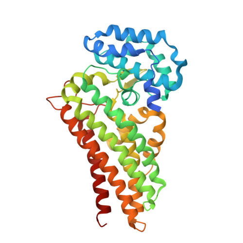
    Pyrrolnitrin, a potent antifungal compound originally discovered in Pseudomonas strains, is biosynthesized through a secondary metabolic pathway involving four key enzymes. Central to this process is PrnB, a heme enzyme that catalyzes the complex transformation of 7-Cl-L-tryptophan. Despite its structural similarity to indoleamine 2,3-dioxygenase and tryptophan 2,3-dioxygenase and its classification within the histidine-ligated heme-dependent aromatic oxygenase superfamily, PrnB has remained relatively unexplored due to the challenges in reconstituting its in vitro activity. In this work, we investigated the interactions of PrnB from different strains with its substrates, substrate analogs, and small molecules using various biophysical and biochemical techniques. Our spectroscopic data reveal that the substrate amino group directly coordinates with the heme in both oxidized and reduced enzyme forms. This binding conformation was further confirmed by X-ray crystallography of enzyme-ligand binary complexes. The amine ligation inhibits H 2 O 2 and CN - from interacting with the ferric heme but does not notably impact NO binding or O 2 activation by the ferrous heme. Stopped-flow spectroscopy showed the formation of heme-based oxidants similar to those reported in indoleamine 2,3-dioxygenase and tryptophan 2,3-dioxygenase when PrnB was exposed to H 2 O 2 or O 2 . However, these intermediates lacked catalytic activity, and PrnB was inactive when coupled with common redox systems under various conditions. This suggests that PrnB operates through a catalytic mechanism distinct from other heme-dependent aromatic oxygenases and most heme enzymes. Our study provides new insights into ligand binding and small-molecule activation mechanisms of PrnB, highlighting its unique functionality and distinguishing it from existing paradigms in heme catalysis.


  • Organizational Affiliation

    Department of Chemistry, University of Georgia, Athens, Georgia, USA.


Macromolecules
Find similar proteins by:  (by identity cutoff)  |  3D Structure
Entity ID: 1
MoleculeChains Sequence LengthOrganismDetailsImage
PrnB
A, B
370Flavobacteriales bacteriumMutation(s): 0 
Entity Groups  
Sequence Clusters30% Identity50% Identity70% Identity90% Identity95% Identity100% Identity
Sequence Annotations
Expand
  • Reference Sequence
Small Molecules
Ligands 3 Unique
IDChains Name / Formula / InChI Key2D Diagram3D Interactions
HEM (Subject of Investigation/LOI)
Query on HEM

Download Ideal Coordinates CCD File 
I [auth A],
K [auth B]
PROTOPORPHYRIN IX CONTAINING FE
C34 H32 Fe N4 O4
KABFMIBPWCXCRK-RGGAHWMASA-L
TSS (Subject of Investigation/LOI)
Query on TSS

Download Ideal Coordinates CCD File 
C [auth A],
J [auth B]
2-(1H-INDOL-3-YL)ETHANAMINE
C10 H12 N2
APJYDQYYACXCRM-UHFFFAOYSA-N
GOL
Query on GOL

Download Ideal Coordinates CCD File 
D [auth A]
E [auth A]
F [auth A]
G [auth A]
H [auth A]
D [auth A],
E [auth A],
F [auth A],
G [auth A],
H [auth A],
L [auth B],
M [auth B],
N [auth B]
GLYCEROL
C3 H8 O3
PEDCQBHIVMGVHV-UHFFFAOYSA-N
Experimental Data & Validation

Experimental Data

  • Method: X-RAY DIFFRACTION
  • Resolution: 2.25 Å
  • R-Value Free:  0.260 (Depositor), 0.258 (DCC) 
  • R-Value Work:  0.206 (Depositor), 0.205 (DCC) 
  • R-Value Observed: 0.208 (Depositor) 
Space Group: C 1 2 1
Unit Cell:
Length ( Å )Angle ( ˚ )
a = 140.744α = 90
b = 122.358β = 94.62
c = 66.474γ = 90
Software Package:
Software NamePurpose
PHENIXrefinement
SCALEPACKdata scaling
DENZOdata reduction
PHENIXphasing

Structure Validation

View Full Validation Report


Ligand Structure Quality Assessment 


Entry History & Funding Information

Deposition Data


Funding OrganizationLocationGrant Number
National Institutes of Health/National Institute of General Medical Sciences (NIH/NIGMS)United StatesR35GM147510

Revision History  (Full details and data files)

  • Version 1.0: 2025-04-16
    Type: Initial release