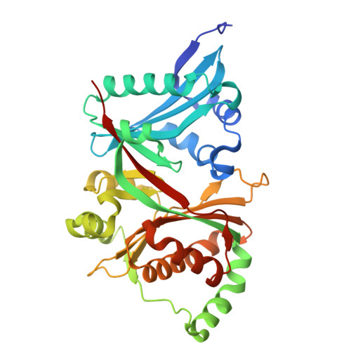
The molecular basis for acetylhistidine synthesis by HisAT/NAT16.
Myllykoski, M., Lundekvam, M., Osberg, C., Nilsen, S.S., Arnesen, T.(2025) Nat Commun 16: 5960-5960
- PubMed: 40595645
- DOI: https://doi.org/10.1038/s41467-025-61145-x
- Primary Citation of Related Structures:
9EMD, 9EMO, 9EMP, 9EMT, 9EN3 - PubMed Abstract:
Acetylhistidine has been detected in human blood, but its origin and function are not known. It is formed when the acetyl group of acetyl-CoA is transferred to the α-amino group of histidine. Here we identify the intracellular NAT16 as the human histidine acetyltransferase (HisAT) responsible for histidine acetylation in vitro and in vivo. A NAT16 variant (p.Phe63Ser) present in over 5% of the population was previously found to correlate with reduced plasma levels of acetylhistidine and increased risk of kidney disease. Our biochemical analysis of HisAT/NAT16 Phe63Ser shows reduced affinity for Histidine supporting a model where this variant has less acetylhistidine catalysis leading to lower blood level of acetylhistidine. We find that HisAT adopts a double-GNAT (Gcn5-related N-Acetyltransferase) fold where the N-terminal domain binds acetyl-CoA and with distinct active site conformation allowing the binding of histidine in between the two domains. We detect similar structures from across living organisms and find that the HisAT structure is conserved in several archaeal and bacterial species. In sum, NAT16 is the human histidine acetyltransferase utilizing a rare double-GNAT structure to steer plasma acetylhistidine levels with potential impact for kidney function.
- Department of Biomedicine, University of Bergen, Bergen, Norway. matti.myllykoski@uib.no.
Organizational Affiliation:


