


Universal Serial electron diffraction for high quality protein structures
Hofer, G., Wang, L., Pacoste, L., Hager, P., Fonjallaz, A., Williams, L., Worrall, J., Steiner, R., Xu, H., Zou, X.To be published.
Experimental Data Snapshot
Starting Model: experimental
View more details
wwPDB Validation 3D Report Full Report

Entity ID: 1 | |||||
|---|---|---|---|---|---|
| Molecule | Chains | Sequence Length | Organism | Details | Image |
| Deferrochelatase | 373 | Streptomyces lividans | Mutation(s): 0 Gene Names: SLI_2602 EC: 1.11.1 | 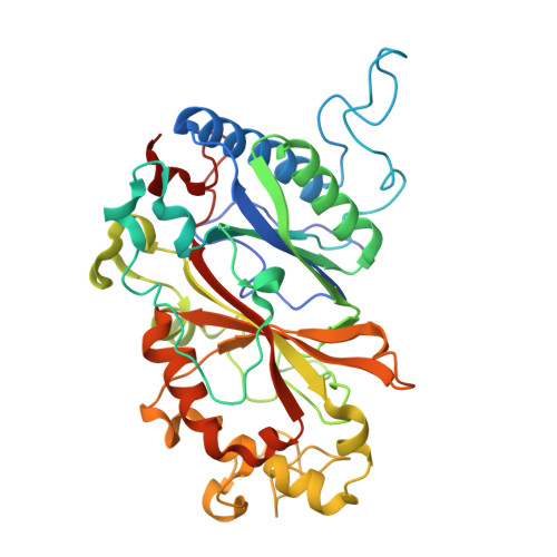 | |
UniProt | |||||
Find proteins for A0ABF7PG09 (Streptomyces lividans TK24) Explore A0ABF7PG09 Go to UniProtKB: A0ABF7PG09 | |||||
Entity Groups | |||||
| Sequence Clusters | 30% Identity50% Identity70% Identity90% Identity95% Identity100% Identity | ||||
| UniProt Group | A0ABF7PG09 | ||||
Sequence AnnotationsExpand | |||||
| |||||
| Ligands 1 Unique | |||||
|---|---|---|---|---|---|
| ID | Chains | Name / Formula / InChI Key | 2D Diagram | 3D Interactions | |
| HEM (Subject of Investigation/LOI) Query on HEM | C [auth A], D [auth B] | PROTOPORPHYRIN IX CONTAINING FE C34 H32 Fe N4 O4 KABFMIBPWCXCRK-RGGAHWMASA-L |  | ||
| Length ( Å ) | Angle ( ˚ ) |
|---|---|
| a = 72.77 | α = 90 |
| b = 67.4 | β = 105.8 |
| c = 73.63 | γ = 90 |
| Task | Software Package | Version |
|---|---|---|
| RECONSTRUCTION | PHENIX | |
| MODEL REFINEMENT | PHENIX |
| Funding Organization | Location | Grant Number |
|---|---|---|
| Knut and Alice Wallenberg Foundation | Sweden | -- |
| Swedish Research Council | Sweden | -- |


