9FZJ | pdb_00009fzj

A new crystal structure of the hPXR-LBD in complex with SR12813 (P43212 form)


Experimental Data Snapshot

  • Method: X-RAY DIFFRACTION
  • Resolution: 1.60 Å
  • R-Value Free: 
    0.209 (Depositor), 0.205 (DCC) 
  • R-Value Work: 
    0.182 (Depositor), 0.180 (DCC) 
  • R-Value Observed: 
    0.183 (Depositor) 

Starting Model: experimental
View more details

wwPDB Validation   3D Report Full Report


Ligand Structure Quality Assessment 


This is version 1.1 of the entry. See complete history


Literature

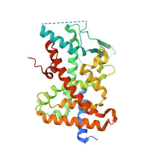
A two-in-one expression construct for biophysical and structural studies of the human pregnane X receptor ligand-binding domain, a pharmaceutical and environmental target.

Carivenc, C.Laconde, G.Blanc, P.Amblard, M.Bourguet, W.Delfosse, V.

(2025) Acta Crystallogr F Struct Biol Commun 81: 85-94

  • DOI: https://doi.org/10.1107/S2053230X2500069X
  • Primary Citation of Related Structures:  
    9FZG, 9FZH, 9FZI, 9FZJ

  • PubMed Abstract: 

    The ligand-binding domain (LBD) of the human nuclear receptor pregnane X receptor (PXR) is known to crystallize in two different crystal forms, P2 1 2 1 2 1 or P4 3 2 1 2, depending on the construct and the strategy used for protein production, as well as the presence or absence of the coactivator-derived peptide SRC-1. In order to facilitate biophysical and structural studies, a versatile construct was designed that allows access to both forms. This was achieved by introducing a thrombin cleavage site between the PXR LBD and the SRC-1 peptide fused to its C-terminus. Here, we describe the expression, purification and crystallization processes of this novel construct and report two new structures of PXR LBD that were obtained thanks to this strategy.


  • Organizational Affiliation

    Centre de Biologie Structurale (CBS), Univ. Montpellier, INSERM, CNRS, Montpellier, France.


Macromolecules
Find similar proteins by:  (by identity cutoff)  |  3D Structure
Entity ID: 1
MoleculeChains Sequence LengthOrganismDetailsImage
Nuclear receptor subfamily 1 group I member 2320Homo sapiensMutation(s): 0 
Gene Names: NR1I2PXR
UniProt & NIH Common Fund Data Resources
Find proteins for O75469 (Homo sapiens)
Explore O75469 
Go to UniProtKB:  O75469
PHAROS:  O75469
GTEx:  ENSG00000144852 
Entity Groups  
Sequence Clusters30% Identity50% Identity70% Identity90% Identity95% Identity100% Identity
UniProt GroupO75469
Sequence Annotations
Expand
  • Reference Sequence
Small Molecules
Ligands 4 Unique
IDChains Name / Formula / InChI Key2D Diagram3D Interactions
SRL (Subject of Investigation/LOI)
Query on SRL

Download Ideal Coordinates CCD File 
F [auth A][2-(3,5-DI-TERT-BUTYL-4-HYDROXY-PHENYL)-1-(DIETHOXY-PHOSPHORYL)-VINYL]-PHOSPHONIC ACID DIETHLYL ESTER
C24 H42 O7 P2
YQLJDECYQDRSBI-UHFFFAOYSA-N
GOL
Query on GOL

Download Ideal Coordinates CCD File 
D [auth A]GLYCEROL
C3 H8 O3
PEDCQBHIVMGVHV-UHFFFAOYSA-N
EDO
Query on EDO

Download Ideal Coordinates CCD File 
B [auth A],
C [auth A]
1,2-ETHANEDIOL
C2 H6 O2
LYCAIKOWRPUZTN-UHFFFAOYSA-N
IPA
Query on IPA

Download Ideal Coordinates CCD File 
E [auth A]ISOPROPYL ALCOHOL
C3 H8 O
KFZMGEQAYNKOFK-UHFFFAOYSA-N
Experimental Data & Validation

Experimental Data

  • Method: X-RAY DIFFRACTION
  • Resolution: 1.60 Å
  • R-Value Free:  0.209 (Depositor), 0.205 (DCC) 
  • R-Value Work:  0.182 (Depositor), 0.180 (DCC) 
  • R-Value Observed: 0.183 (Depositor) 
Space Group: P 43 21 2
Unit Cell:
Length ( Å )Angle ( ˚ )
a = 91.52α = 90
b = 91.52β = 90
c = 85.7γ = 90
Software Package:
Software NamePurpose
PHENIXrefinement
XDSdata reduction
XSCALEdata scaling
PHENIXphasing

Structure Validation

View Full Validation Report


Ligand Structure Quality Assessment 


Entry History & Funding Information

Deposition Data


Funding OrganizationLocationGrant Number
Agence Nationale de la Recherche (ANR)France--

Revision History  (Full details and data files)

  • Version 1.0: 2025-02-19
    Type: Initial release
  • Version 1.1: 2025-03-12
    Changes: Database references