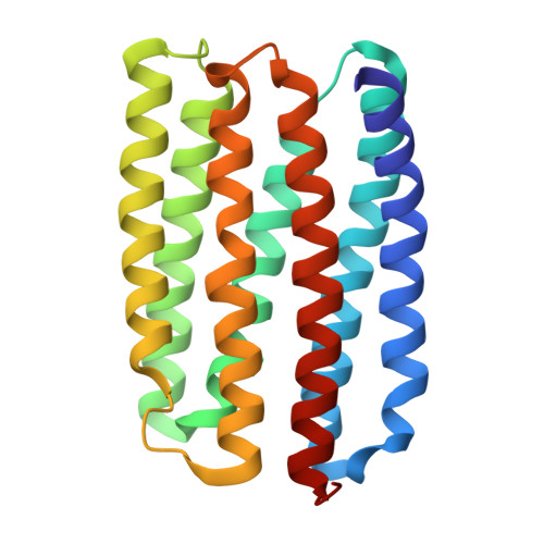
9G16 | pdb_00009g16

Crystal structure of marine actinobacteria clade rhodopsin (MAR) in the O state obtained by cryotrapping


Experimental Data Snapshot

  • Method: X-RAY DIFFRACTION
  • Resolution: 1.51 Å
  • R-Value Free: 
    0.208 (Depositor), 0.208 (DCC) 
  • R-Value Work: 
    0.175 (Depositor), 0.175 (DCC) 
  • R-Value Observed: 
    0.177 (Depositor) 

Starting Model: experimental
View more details

wwPDB Validation   3D Report Full Report


Ligand Structure Quality Assessment 


This is version 1.1 of the entry. See complete history


Literature

Proteorhodopsin insights into the molecular mechanism of vectorial proton transport.

Bukhdruker, S.Gushchin, I.Shevchenko, V.Kovalev, K.Polovinkin, V.Tsybrov, F.Astashkin, R.Alekseev, A.Mikhaylov, A.Bukhalovich, S.Bratanov, D.Ryzhykau, Y.Kuklina, D.Caramello, N.Rokitskaya, T.Antonenko, Y.Rulev, M.Stoev, C.Zabelskii, D.Round, E.Rogachev, A.Borshchevskiy, V.Ghai, R.Bourenkov, G.Zeghouf, M.Cherfils, J.Engelhard, M.Chizhov, I.Rodriguez-Valera, F.Bamberg, E.Gordeliy, V.

(2025) Sci Adv 11: eadu5303-eadu5303

  • DOI: https://doi.org/10.1126/sciadv.adu5303
  • Primary Citation of Related Structures:  
    7AVN, 7AVP, 8RSO, 8RSP, 8RSQ, 8RSR, 8RSS, 9G15, 9G16

  • PubMed Abstract: 

    Bacterial proton pumps, proteorhodopsins (PRs), are a major group of light-driven membrane proteins found in marine bacteria. They are functionally and structurally distinct from archaeal and eukaryotic proton pumps. To elucidate the proton transfer mechanism by PRs and understand the differences to nonbacterial pumps on a molecular level, high-resolution structures of PRs' functional states are needed. In this work, we have determined atomic-resolution structures of MAR, a PR from marine actinobacteria, in various functional states, notably the challenging late O intermediate state. These data and information from recent atomic-resolution structures on an archaeal outward proton pump bacteriorhodopsin and bacterial inward proton pump xenorhodopsin allow for deducing key universal elements for light-driven proton pumping. First, long hydrogen-bonded chains characterize proton pathways. Second, short hydrogen bonds allow proton storage and inhibit their backflow. Last, the retinal Schiff base is the active proton donor and acceptor to and from hydrogen-bonded chains.


  • Organizational Affiliation

    Research Center for Molecular Mechanisms of Aging and Age-Related Diseases, Moscow Institute of Physics and Technology, 141700 Dolgoprudny, Russia.


Macromolecules
Find similar proteins by:  (by identity cutoff)  |  3D Structure
Entity ID: 1
MoleculeChains Sequence LengthOrganismDetailsImage
Bacteriorhodopsin220marine Actinobacteria cladeMutation(s): 0 
Membrane Entity: Yes 
UniProt
Find proteins for S5DM51 (Candidatus Actinomarina minuta)
Explore S5DM51 
Go to UniProtKB:  S5DM51
Entity Groups  
Sequence Clusters30% Identity50% Identity70% Identity90% Identity95% Identity100% Identity
UniProt GroupS5DM51
Sequence Annotations
Expand
  • Reference Sequence
Small Molecules
Ligands 3 Unique
IDChains Name / Formula / InChI Key2D Diagram3D Interactions
RET (Subject of Investigation/LOI)
Query on RET

Download Ideal Coordinates CCD File 
P [auth A]RETINAL
C20 H28 O
NCYCYZXNIZJOKI-OVSJKPMPSA-N
LFA
Query on LFA

Download Ideal Coordinates CCD File 
E [auth A]
F [auth A]
G [auth A]
H [auth A]
I [auth A]
E [auth A],
F [auth A],
G [auth A],
H [auth A],
I [auth A],
J [auth A],
K [auth A],
L [auth A],
M [auth A],
N [auth A],
O [auth A]
EICOSANE
C20 H42
CBFCDTFDPHXCNY-UHFFFAOYSA-N
OLA
Query on OLA

Download Ideal Coordinates CCD File 
B [auth A],
C [auth A],
D [auth A]
OLEIC ACID
C18 H34 O2
ZQPPMHVWECSIRJ-KTKRTIGZSA-N
Experimental Data & Validation

Experimental Data

  • Method: X-RAY DIFFRACTION
  • Resolution: 1.51 Å
  • R-Value Free:  0.208 (Depositor), 0.208 (DCC) 
  • R-Value Work:  0.175 (Depositor), 0.175 (DCC) 
  • R-Value Observed: 0.177 (Depositor) 
Space Group: P 1 2 1
Unit Cell:
Length ( Å )Angle ( ˚ )
a = 50.537α = 90
b = 40.311β = 101.25
c = 60.299γ = 90
Software Package:
Software NamePurpose
XDSdata reduction
STARANISOdata scaling
PHASERphasing
PHENIXrefinement

Structure Validation

View Full Validation Report


Ligand Structure Quality Assessment 


Entry History & Funding Information

Deposition Data


Funding OrganizationLocationGrant Number
Agence Nationale de la Recherche (ANR)France--

Revision History  (Full details and data files)

  • Version 1.0: 2025-04-02
    Type: Initial release
  • Version 1.1: 2025-04-23
    Changes: Database references, Structure summary